Loading
PDBj
MenuPDBj@FacebookPDBj@TwitterPDBj@YouTubewwPDB FoundationwwPDB
RCSB PDBPDBeBMRBAdv. SearchSearch help
Search by PDB author
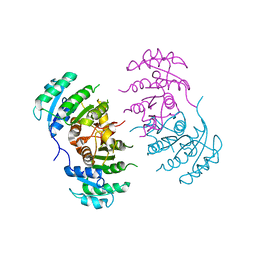
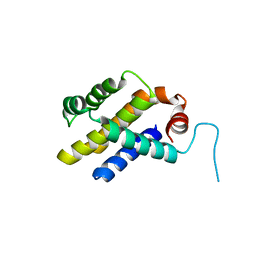
3IIG
DownloadVisualize
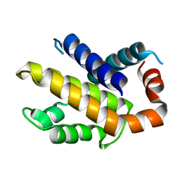
BU of 3iig by Molmil
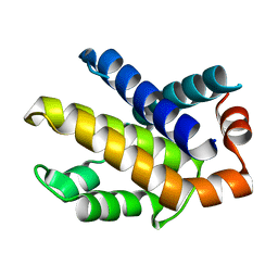
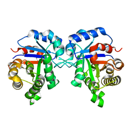
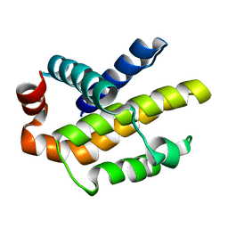
Crystal structure of mouse Bcl-xl mutant (F105A) at pH 5.0
Descriptor: Bcl-2-like protein 1
Authors:Priyadarshi, A, Hwang, K.Y.
Deposit date:2009-08-01
Release date:2010-04-14
Last modified:2023-11-01
Method:X-RAY DIFFRACTION (2.3 Å)
Cite:Structural insights into mouse anti-apoptotic Bcl-xl reveal affinity for Beclin 1 and gossypol.
Biochem.Biophys.Res.Commun., 394, 2010
3ILB
DownloadVisualize
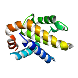
BU of 3ilb by Molmil
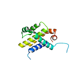
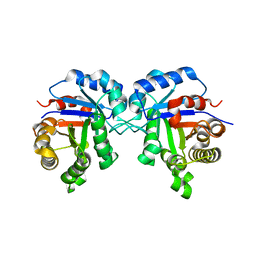
Crystal structure of mouse Bcl-xl mutant (R139A) at pH 6.0
Descriptor: Bcl-2-like protein 1
Authors:Priyadarshi, A, Hwang, K.Y.
Deposit date:2009-08-07
Release date:2010-04-14
Last modified:2023-11-01
Method:X-RAY DIFFRACTION (2.38 Å)
Cite:Structural insights into mouse anti-apoptotic Bcl-xl reveal affinity for Beclin 1 and gossypol.
Biochem.Biophys.Res.Commun., 394, 2010
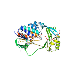
7E43
DownloadVisualize
BU of 7e43 by Molmil
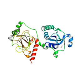
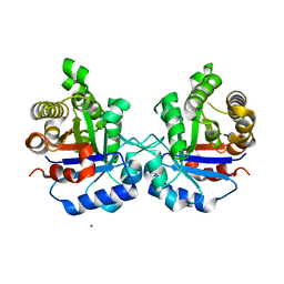
Structural insights into a bifunctional peptide methionine sulfoxide reductase MsrA/B fusion protein from Helicobacter pylori
Descriptor: ACETATE ION, Peptide methionine sulfoxide reductase MsrA/MsrB
Authors:Kim, S, Lee, K, Hwang, K.Y.
Deposit date:2021-02-10
Release date:2021-04-21
Last modified:2023-11-29
Method:X-RAY DIFFRACTION (2.2 Å)
Cite:Structural Insights into a Bifunctional Peptide Methionine Sulfoxide Reductase MsrA/B Fusion Protein from Helicobacter pylori .
Antioxidants (Basel), 10, 2021
3IHD
DownloadVisualize
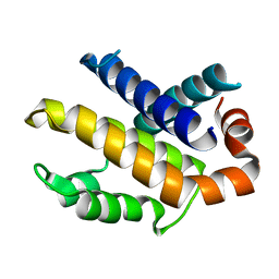
BU of 3ihd by Molmil
Crystal structure of mouse Bcl-xl mutant (Y101A) at pH 5.0
Descriptor: Bcl-2-like protein 1
Authors:Priyadarshi, A, Hwang, K.Y.
Deposit date:2009-07-30
Release date:2010-04-14
Last modified:2023-11-01
Method:X-RAY DIFFRACTION (1.88 Å)
Cite:Structural insights into mouse anti-apoptotic Bcl-xl reveal affinity for Beclin 1 and gossypol.
Biochem.Biophys.Res.Commun., 394, 2010
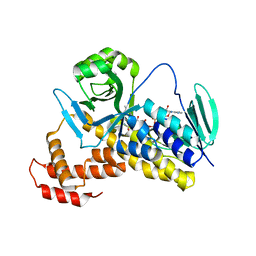
3HWW
DownloadVisualize
BU of 3hww by Molmil
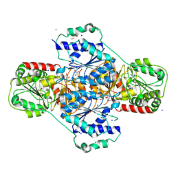
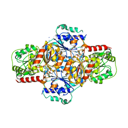
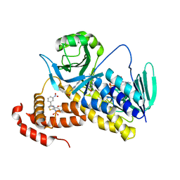
Crystal structure of menaquinone synthesis protein MenD from E. coli in complex with oxoglutarate
Descriptor: 2-OXOGLUTARIC ACID, 2-succinyl-5-enolpyruvyl-6-hydroxy-3-cyclohexene-1-carboxylate synthase, CHLORIDE ION, ...
Authors:Priyadarshi, A, Hwang, K.Y.
Deposit date:2009-06-19
Release date:2009-11-03
Last modified:2023-11-01
Method:X-RAY DIFFRACTION (1.95 Å)
Cite:Structural and functional analysis of Vitamin K2 synthesis protein MenD.
Biochem.Biophys.Res.Commun., 388, 2009
3IHF
DownloadVisualize
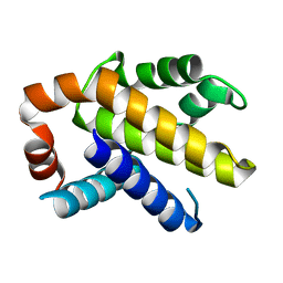
BU of 3ihf by Molmil
Crystal structure of mouse Bcl-xl mutant (R139A) at pH 5.0
Descriptor: Bcl-2-like protein 1
Authors:Priyadarshi, A, Hwang, K.Y.
Deposit date:2009-07-30
Release date:2010-04-14
Last modified:2023-11-01
Method:X-RAY DIFFRACTION (2.28 Å)
Cite:Structural insights into mouse anti-apoptotic Bcl-xl reveal affinity for Beclin 1 and gossypol.
Biochem.Biophys.Res.Commun., 394, 2010
3IIH
DownloadVisualize
BU of 3iih by Molmil
Crystal structure of mouse Bcl-xl (wt) at pH 6.0
Descriptor: Bcl-2-like protein 1
Authors:Priyadarshi, A, Hwang, K.Y.
Deposit date:2009-08-01
Release date:2010-04-14
Last modified:2023-11-01
Method:X-RAY DIFFRACTION (2.75 Å)
Cite:Structural insights into mouse anti-apoptotic Bcl-xl reveal affinity for Beclin 1 and gossypol.
Biochem.Biophys.Res.Commun., 394, 2010
3IHE
DownloadVisualize
BU of 3ihe by Molmil
Crystal structure of mouse Bcl-xl mutant (F105A) at pH 6.0
Descriptor: Bcl-2-like protein 1
Authors:Priyadarshi, A, Hwang, K.Y.
Deposit date:2009-07-30
Release date:2010-04-14
Last modified:2023-11-01
Method:X-RAY DIFFRACTION (3 Å)
Cite:Structural insights into mouse anti-apoptotic Bcl-xl reveal affinity for Beclin 1 and gossypol.
Biochem.Biophys.Res.Commun., 394, 2010
3ILC
DownloadVisualize
BU of 3ilc by Molmil
Crystal structure of mouse Bcl-xl mutant (Y101A) at pH 6.0
Descriptor: Bcl-2-like protein 1
Authors:Priyadarshi, A, Hwang, K.Y.
Deposit date:2009-08-07
Release date:2010-04-14
Last modified:2023-11-01
Method:X-RAY DIFFRACTION (1.64 Å)
Cite:Structural insights into mouse anti-apoptotic Bcl-xl reveal affinity for Beclin 1 and gossypol.
Biochem.Biophys.Res.Commun., 2010
3IHC
DownloadVisualize
BU of 3ihc by Molmil
Crystal structure of mouse Bcl-xl (wt) at pH 5.0
Descriptor: Bcl-2-like protein 1
Authors:Priyadarshi, A, Hwang, K.Y.
Deposit date:2009-07-30
Release date:2010-04-14
Last modified:2023-11-01
Method:X-RAY DIFFRACTION (1.85 Å)
Cite:Structural insights into mouse anti-apoptotic Bcl-xl reveal affinity for Beclin 1 and gossypol.
Biochem.Biophys.Res.Commun., 394, 2010
3GNT
DownloadVisualize
BU of 3gnt by Molmil
Crystal Structure of the Staphylococcus aureus Enoyl-Acyl Carrier Protein Reductase (FabI) in apo form (two molecules in AU)
Descriptor: Enoyl-[acyl-carrier-protein] reductase [NADH]
Authors:Priyadarshi, A, Hwang, K.Y.
Deposit date:2009-03-18
Release date:2009-10-20
Last modified:2023-11-01
Method:X-RAY DIFFRACTION (2.75 Å)
Cite:Structural insights into Staphylococcus aureus enoyl-ACP reductase (FabI), in complex with NADP and triclosan.
Proteins, 78, 2010
7CPZ
DownloadVisualize
BU of 7cpz by Molmil
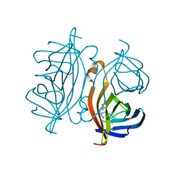
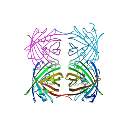
Crystal structure of Streptoavidin-C1 from Streptomyces cinamonensis
Descriptor: BIOTIN, Mature Streptoavidin-C1
Authors:Jeon, B.J, Kim, S, Lee, J.-H, Kim, M.S, Hwang, K.Y.
Deposit date:2020-08-08
Release date:2021-07-14
Last modified:2023-11-29
Method:X-RAY DIFFRACTION (2.5 Å)
Cite:Insights into the structure of mature streptavidin C1 from Streptomyces cinnamonensis reveal the self-binding of the extension C-terminal peptide to biotin-binding sites.
Iucrj, 8, 2021
7CQ0
DownloadVisualize
BU of 7cq0 by Molmil
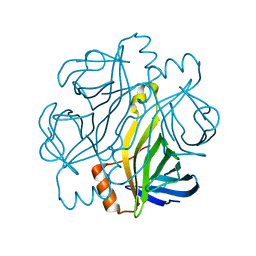
Crystal structure of Streptoavidin-C1 from Streptomyces cinamonensis
Descriptor: Mature Streptoavidin-C1
Authors:Jeon, B.J, Kim, S, Lee, J.-H, Kim, M.S, Hwang, K.Y.
Deposit date:2020-08-08
Release date:2021-07-14
Last modified:2023-11-29
Method:X-RAY DIFFRACTION (2.03 Å)
Cite:Insights into the structure of mature streptavidin C1 from Streptomyces cinnamonensis reveal the self-binding of the extension C-terminal peptide to biotin-binding sites.
Iucrj, 8, 2021
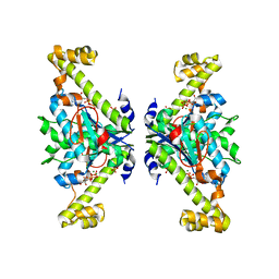
3HWX
DownloadVisualize
BU of 3hwx by Molmil
Crystal structure of menaquinone synthesis protein MenD from E. coli in complex with ThDP
Descriptor: 2-succinyl-5-enolpyruvyl-6-hydroxy-3-cyclohexene-1-carboxylate synthase, GLYCEROL, MAGNESIUM ION, ...
Authors:Priyadarshi, A, Hwang, K.Y.
Deposit date:2009-06-19
Release date:2009-11-03
Last modified:2023-11-01
Method:X-RAY DIFFRACTION (2.6 Å)
Cite:Structural and functional analysis of Vitamin K2 synthesis protein MenD.
Biochem.Biophys.Res.Commun., 388, 2009
5X68
DownloadVisualize
BU of 5x68 by Molmil
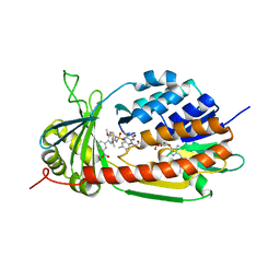
Crystal Structure of Human KMO
Descriptor: FLAVIN-ADENINE DINUCLEOTIDE, Kynurenine 3-monooxygenase
Authors:Kim, H.T, Hwang, K.Y.
Deposit date:2017-02-21
Release date:2018-02-21
Last modified:2018-05-02
Method:X-RAY DIFFRACTION (2.1 Å)
Cite:Structural Basis for Inhibitor-Induced Hydrogen Peroxide Production by Kynurenine 3-Monooxygenase
Cell Chem Biol, 25, 2018
5X6P
DownloadVisualize
BU of 5x6p by Molmil
Crystal structure of Pseudomonas fluorescens KMO
Descriptor: FLAVIN-ADENINE DINUCLEOTIDE, Kynurenine 3-monooxygenase
Authors:Kim, H.T, Hwang, K.Y.
Deposit date:2017-02-22
Release date:2018-02-21
Last modified:2023-11-22
Method:X-RAY DIFFRACTION (1.78 Å)
Cite:Structural Basis for Inhibitor-Induced Hydrogen Peroxide Production by Kynurenine 3-Monooxygenase
Cell Chem Biol, 25, 2018
5XOX
DownloadVisualize
BU of 5xox by Molmil
Crystal structure of tRNA(His) guanylyltranserase from Saccharomyces cerevisiae
Descriptor: GUANOSINE-5'-TRIPHOSPHATE, MAGNESIUM ION, PHOSPHATE ION, ...
Authors:Lee, K, Lee, E.H, Son, J, Hwang, K.Y.
Deposit date:2017-05-31
Release date:2017-07-12
Last modified:2023-11-22
Method:X-RAY DIFFRACTION (3 Å)
Cite:Crystal structure of tRNA(His) guanylyltransferase from Saccharomyces cerevisiae
Biochem. Biophys. Res. Commun., 490, 2017
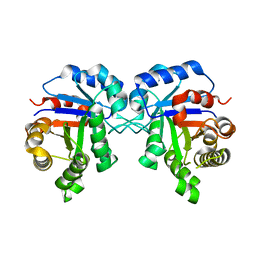
5ZGA
DownloadVisualize
BU of 5zga by Molmil
Crystal Structure of Triosephosphate isomerase SAD deletion and N115A mutant from Opisthorchis viverrini
Descriptor: Triosephosphate isomerase
Authors:Son, J, Kim, S, Kim, S.E, Lee, H, Lee, M.R, Hwang, K.Y.
Deposit date:2018-03-08
Release date:2018-10-24
Last modified:2023-11-22
Method:X-RAY DIFFRACTION (1.793 Å)
Cite:Structural Analysis of an Epitope Candidate of Triosephosphate Isomerase in Opisthorchis viverrini.
Sci Rep, 8, 2018
5ZG5
DownloadVisualize
BU of 5zg5 by Molmil
Crystal Structure of Triosephosphate isomerase SADsubAAA mutant from Opisthorchis viverrini
Descriptor: Triosephosphate isomerase
Authors:Son, J, Kim, S, Kim, S.E, Lee, H, Lee, M.R, Hwang, K.Y.
Deposit date:2018-03-07
Release date:2018-10-24
Last modified:2023-11-22
Method:X-RAY DIFFRACTION (1.597 Å)
Cite:Structural Analysis of an Epitope Candidate of Triosephosphate Isomerase in Opisthorchis viverrini.
Sci Rep, 8, 2018
5ZG4
DownloadVisualize
BU of 5zg4 by Molmil
Crystal Structure of Triosephosphate isomerase SAD deletion mutant from Opisthorchis viverrini
Descriptor: Triosephosphate isomerase
Authors:Son, J, Kim, S, Kim, S.E, Lee, H, Lee, M.R, Hwang, K.Y.
Deposit date:2018-03-07
Release date:2018-10-24
Last modified:2023-11-22
Method:X-RAY DIFFRACTION (1.746 Å)
Cite:Structural Analysis of an Epitope Candidate of Triosephosphate Isomerase in Opisthorchis viverrini.
Sci Rep, 8, 2018
5ZFX
DownloadVisualize
BU of 5zfx by Molmil
Crystal Structure of Triosephosphate isomerase from Opisthorchis viverrini
Descriptor: MAGNESIUM ION, Triosephosphate isomerase
Authors:Son, J, Kim, S, Kim, S.E, Lee, H, Lee, M.R, Hwang, K.Y.
Deposit date:2018-03-07
Release date:2018-10-24
Last modified:2023-11-22
Method:X-RAY DIFFRACTION (1.751 Å)
Cite:Structural Analysis of an Epitope Candidate of Triosephosphate Isomerase in Opisthorchis viverrini.
Sci Rep, 8, 2018
6AA7
DownloadVisualize
BU of 6aa7 by Molmil
Fluorescent protein from Acropora digitifera
Descriptor: Fluorescent protein
Authors:Kim, S.E, Hwang, K.Y, Nam, K.H.
Deposit date:2018-07-17
Release date:2018-08-22
Last modified:2024-10-09
Method:X-RAY DIFFRACTION (1.8 Å)
Cite:Spectral and structural analysis of a red fluorescent protein from Acropora digitifera.
Protein Sci., 28, 2019
5X6Q
DownloadVisualize
BU of 5x6q by Molmil
Crystal structure of Pseudomonas fluorescens KMO in complex with Ro 61-8048
Descriptor: 3,4-dimethoxy-N-[4-(3-nitrophenyl)-1,3-thiazol-2-yl]benzenesulfonamide, FLAVIN-ADENINE DINUCLEOTIDE, Kynurenine 3-monooxygenase
Authors:Kim, H.T, Hwang, K.Y.
Deposit date:2017-02-23
Release date:2018-02-21
Last modified:2023-11-22
Method:X-RAY DIFFRACTION (1.897 Å)
Cite:Structural Basis for Inhibitor-Induced Hydrogen Peroxide Production by Kynurenine 3-Monooxygenase
Cell Chem Biol, 25, 2018
5X6R
DownloadVisualize
BU of 5x6r by Molmil
Crystal structure of Saccharomyces cerevisiae KMO in complex with Ro 61-8048
Descriptor: 3,4-dimethoxy-N-[4-(3-nitrophenyl)-1,3-thiazol-2-yl]benzenesulfonamide, FLAVIN-ADENINE DINUCLEOTIDE, Kynurenine 3-monooxygenase
Authors:Kim, H.T, Hwang, K.Y.
Deposit date:2017-02-23
Release date:2018-02-21
Last modified:2023-11-22
Method:X-RAY DIFFRACTION (1.911 Å)
Cite:Structural Basis for Inhibitor-Induced Hydrogen Peroxide Production by Kynurenine 3-Monooxygenase
Cell Chem Biol, 25, 2018
5IZV
DownloadVisualize
BU of 5izv by Molmil
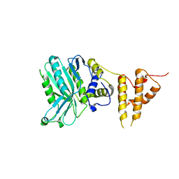
Crystal structure of the legionella pneumophila effector protein RavZ - F222
Descriptor: Uncharacterized protein RavZ
Authors:Kwon, D.H, Kim, L, Kim, B.-W, Hong, S.B, Song, H.K.
Deposit date:2016-03-26
Release date:2016-11-09
Last modified:2024-03-20
Method:X-RAY DIFFRACTION (2.814 Å)
Cite:The 1:2 complex between RavZ and LC3 reveals a mechanism for deconjugation of LC3 on the phagophore membrane
Autophagy, 13, 2017

226707

PDB entries from 2024-10-30

PDB statisticsPDBj update infoContact PDBjnumon