Loading
PDBj
MenuPDBj@FacebookPDBj@TwitterPDBj@YouTubewwPDB FoundationwwPDB
RCSB PDBPDBeBMRBAdv. SearchSearch help
Search by PDB author
6E5G
DownloadVisualize
BU of 6e5g by Molmil
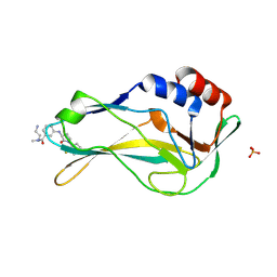
Crystal structure of human TEAD2-Yap binding domain covalently bound to an allosteric regulator
Descriptor: 1-(2-{[3-(trifluoromethyl)phenyl]amino}phenyl)ethan-1-one, Transcriptional enhancer factor TEF-4
Authors:Bum-Erdene, K, Gonzalez-Gutierrez, G, Meroueh, S.O.
Deposit date:2018-07-20
Release date:2019-01-09
Last modified:2024-10-16
Method:X-RAY DIFFRACTION (2.43 Å)
Cite:Small-Molecule Covalent Modification of Conserved Cysteine Leads to Allosteric Inhibition of the TEAD⋅Yap Protein-Protein Interaction.
Cell Chem Biol, 26, 2019
6P0J
DownloadVisualize
BU of 6p0j by Molmil
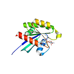
Crystal structure of GDP-bound human RalA
Descriptor: CALCIUM ION, GUANOSINE-5'-DIPHOSPHATE, Ras-related protein Ral-A
Authors:Bum-Erdene, K, Gonzalez-Gutierrez, G, Liu, D, Meroueh, S.O.
Deposit date:2019-05-17
Release date:2020-03-04
Last modified:2023-10-11
Method:X-RAY DIFFRACTION (1.31 Å)
Cite:Small-molecule covalent bond formation at tyrosine creates a binding site and inhibits activation of Ral GTPases.
Proc.Natl.Acad.Sci.USA, 117, 2020
6PHJ
DownloadVisualize
BU of 6phj by Molmil
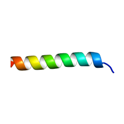
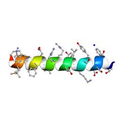
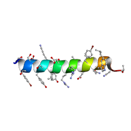
Crystal structure of native glucagon in space group P213 at 1.99 A resolution
Descriptor: Glucagon
Authors:Mroz, P.A, Gonzalez-Gutierrez, G, DiMarchi, R.D.
Deposit date:2019-06-25
Release date:2020-07-01
Last modified:2023-10-11
Method:X-RAY DIFFRACTION (1.99 Å)
Cite:High resolution X-ray structure of glucagon and selected stereo-inversed analogs in novel crystallographic packing arrangement.
To Be Published
6PHO
DownloadVisualize
BU of 6pho by Molmil
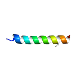
Crystal structure of glucagon analog with selenomethionine substitutions at position 1 and 27 in space group I41 at 1.42 A resolution
Descriptor: Glucagon
Authors:Mroz, P.A, Gonzalez-Gutierrez, G, DiMarchi, R.D.
Deposit date:2019-06-25
Release date:2020-07-01
Last modified:2024-10-09
Method:X-RAY DIFFRACTION (1.42 Å)
Cite:High resolution X-ray structure of glucagon and selected stereo-inversed analogs in novel crystallographic packing arrangement.
To Be Published
6PHK
DownloadVisualize
BU of 6phk by Molmil
Crystal structure of glucagon analog with mono-stereoinversion at position 21 (D-Asp21) in space group I41 at 1.18 A resolution
Descriptor: Glucagon
Authors:Mroz, P.A, Gonzalez-Gutierrez, G, DiMarchi, R.D.
Deposit date:2019-06-25
Release date:2020-07-01
Last modified:2024-10-16
Method:X-RAY DIFFRACTION (1.18 Å)
Cite:High resolution X-ray structure of glucagon and selected stereo-inversed analogs in novel crystallographic packing arrangement.
To Be Published
6PHP
DownloadVisualize
BU of 6php by Molmil
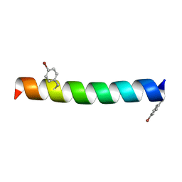
Crystal structure of glucagon analog with 4-bromo-phenylalanine substitutions at position 6 and 22 in space group I41 at 1.65 A resolution
Descriptor: Glucagon
Authors:Mroz, P.A, Gonzalez-Gutierrez, G, DiMarchi, R.D.
Deposit date:2019-06-25
Release date:2020-07-01
Last modified:2023-11-15
Method:X-RAY DIFFRACTION (1.65 Å)
Cite:High resolution X-ray structure of glucagon and selected stereo-inversed analogs in novel crystallographic packing arrangement.
To Be Published
6PHN
DownloadVisualize
BU of 6phn by Molmil
Crystal structure of glucagon analog composed of D-amino acids with mono-stereoinversion at position 23 (L-Val23) in space group I41 at 1.33 A resolution
Descriptor: D-glucagon L-Val23, SODIUM ION
Authors:Mroz, P.A, Gonzalez-Gutierrez, G, DiMarchi, R.D.
Deposit date:2019-06-25
Release date:2020-07-01
Last modified:2024-10-30
Method:X-RAY DIFFRACTION (1.33 Å)
Cite:High resolution X-ray structure of glucagon and selected stereo-inversed analogs in novel crystallographic packing arrangement.
To Be Published
6PHI
DownloadVisualize
BU of 6phi by Molmil
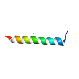
Crystal structure of native glucagon in space group I41 at 1.1 A resolution
Descriptor: CHLORIDE ION, Glucagon
Authors:Mroz, P.A, Gonzalez-Gutierrez, G, DiMarchi, R.D.
Deposit date:2019-06-25
Release date:2020-07-01
Last modified:2023-10-11
Method:X-RAY DIFFRACTION (1.1 Å)
Cite:High resolution X-ray structure of glucagon and selected stereo-inversed analogs in novel crystallographic packing arrangement.
To Be Published
6PHM
DownloadVisualize
BU of 6phm by Molmil
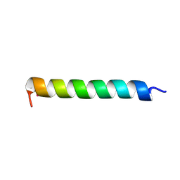
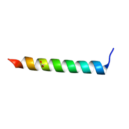
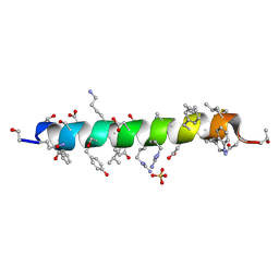
Crystal structure of glucagon analog fully composed of D-amino acids in space group I41 at 1.1 A resolution
Descriptor: D-glucagon, SULFATE ION
Authors:Mroz, P.A, Gonzalez-Gutierrez, G, DiMarchi, R.D.
Deposit date:2019-06-25
Release date:2020-07-01
Last modified:2024-10-23
Method:X-RAY DIFFRACTION (1.1 Å)
Cite:High resolution X-ray structure of glucagon and selected stereo-inversed analogs in novel crystallographic packing arrangement.
To Be Published
6PHL
DownloadVisualize
BU of 6phl by Molmil
Crystal structure of glucagon analog with mono-stereoinversion at position 23 (D-Val23) in space group I41 at 1.44 A resolution
Descriptor: Glucagon
Authors:Mroz, P.A, Gonzalez-Gutierrez, G, DiMarchi, R.D.
Deposit date:2019-06-25
Release date:2020-07-01
Last modified:2024-10-16
Method:X-RAY DIFFRACTION (1.443 Å)
Cite:High resolution X-ray structure of glucagon and selected stereo-inversed analogs in novel crystallographic packing arrangement.
To Be Published
6PHQ
DownloadVisualize
BU of 6phq by Molmil
Crystal structure of glucagon analog fully composed of D-amino acids with 4-bromo-D-phenylalanine substitutions at position 6 and 22 in space group I41 at 1.1 A resolution
Descriptor: D-glucagon D-BrPhe 6,22
Authors:Mroz, P.A, Gonzalez-Gutierrez, G, DiMarchi, R.D.
Deposit date:2019-06-25
Release date:2020-07-01
Last modified:2023-11-15
Method:X-RAY DIFFRACTION (1.32 Å)
Cite:High resolution X-ray structure of glucagon and selected stereo-inversed analogs in novel crystallographic packing arrangement.
To Be Published
7ZBD
DownloadVisualize
BU of 7zbd by Molmil
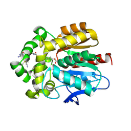
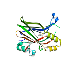
HaloTag with TRaQ-G ligand
Descriptor: (10R)-7-azanyl-N-[2-[2-(6-chloranylhexoxy)ethoxy]ethyl]-2'-cyano-5,5-dimethyl-3-(methylamino)-1'-oxidanylidene-spiro[benzo[b][1]benzosiline-10,3'-isoindole]-5'-carboxamide, CHLORIDE ION, GLYCEROL, ...
Authors:Emmert, S, Rivera-Fuentes, P, Pojer, F, Lau, K.
Deposit date:2022-03-23
Release date:2023-02-01
Last modified:2024-02-07
Method:X-RAY DIFFRACTION (1.68 Å)
Cite:A locally activatable sensor for robust quantification of organellar glutathione.
Nat.Chem., 15, 2023
7ZBA
DownloadVisualize
BU of 7zba by Molmil
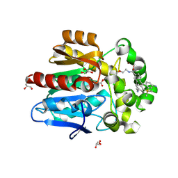
HaloTag with Me-TRaQ-G ligand
Descriptor: 4-(7-azanyl-5,5-dimethyl-3-methylimino-benzo[b][1]benzosilin-10-yl)-N-[2-[2-(6-chloranylhexoxy)ethoxy]ethyl]-3-methyl-benzamide, CHLORIDE ION, GLYCEROL, ...
Authors:Emmert, S, Rivera-Fuentes, P, Pojer, F, Lau, K.
Deposit date:2022-03-23
Release date:2023-02-01
Last modified:2024-10-23
Method:X-RAY DIFFRACTION (1.23 Å)
Cite:A locally activatable sensor for robust quantification of organellar glutathione.
Nat.Chem., 15, 2023
7ZBB
DownloadVisualize
BU of 7zbb by Molmil
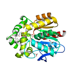
HaloTag with TRaQ-G-ctrl ligand
Descriptor: (E)-[7-azanyl-10-[2-carboxy-5-[2-[2-(6-chloranylhexoxy)ethoxy]ethylcarbamoyl]phenyl]-5,5-dimethyl-benzo[b][1]benzosilin-3-ylidene]-methyl-azanium, CHLORIDE ION, GLYCEROL, ...
Authors:Emmert, S, Rivera-Fuentes, P, Pojer, F, Lau, K.
Deposit date:2022-03-23
Release date:2023-02-01
Last modified:2024-02-07
Method:X-RAY DIFFRACTION (1.95 Å)
Cite:A locally activatable sensor for robust quantification of organellar glutathione.
Nat.Chem., 15, 2023
7Z3T
DownloadVisualize
BU of 7z3t by Molmil
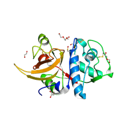
Crystal structure of apo human Cathepsin L
Descriptor: 1,2-ETHANEDIOL, Cathepsin L, DI(HYDROXYETHYL)ETHER, ...
Authors:Reinke, P.Y.A, Falke, S, Lieske, J, Ewert, W, Loboda, J, Rahmani Mashhour, A, Hauser, M, Karnicar, K, Usenik, A, Lindic, N, Lach, M, Boehler, H, Beck, T, Cox, R, Chapman, H.N, Hinrichs, W, Turk, D, Guenther, S, Meents, A.
Deposit date:2022-03-02
Release date:2023-03-22
Last modified:2024-10-16
Method:X-RAY DIFFRACTION (1.6 Å)
Cite:Calpeptin is a potent cathepsin inhibitor and drug candidate for SARS-CoV-2 infections.
Commun Biol, 6, 2023
7YOO
DownloadVisualize
BU of 7yoo by Molmil
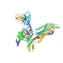
Complex structure of Neuropeptide Y Y2 receptor in complex with NPY and Gi
Descriptor: Guanine nucleotide-binding protein G(I)/G(S)/G(O) subunit gamma-2, Guanine nucleotide-binding protein G(I)/G(S)/G(T) subunit beta-1, Guanine nucleotide-binding protein G(i) subunit alpha-1, ...
Authors:Kang, H, Park, C, Kim, J, Choi, H.-J.
Deposit date:2022-08-01
Release date:2023-03-22
Method:ELECTRON MICROSCOPY (3.11 Å)
Cite:Structural basis for Y2 receptor-mediated neuropeptide Y and peptide YY signaling.
Structure, 31, 2023
7ZGE
DownloadVisualize
BU of 7zge by Molmil
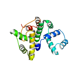
BrxA, BREX phage defence protein
Descriptor: BrxA, BREX phage defence protein
Authors:Picton, D.M, Beck, I.N, Blower, T.R.
Deposit date:2022-04-03
Release date:2022-06-22
Last modified:2024-03-20
Method:X-RAY DIFFRACTION (2.09 Å)
Cite:Crystal structure of the anti-CRISPR repressor Aca2.
J.Struct.Biol., 213, 2021
6PW8
DownloadVisualize
BU of 6pw8 by Molmil
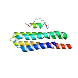
Hydrocarbon-Stapled Paxillin Peptide Bound to the Focal Adhesion Targeting (FAT) Domain of the Focal Adhesion Kinase (FAK)
Descriptor: CHLORIDE ION, Focal adhesion kinase 1, SP3, ...
Authors:Thifault, D.G, Fromme, P, Martin-Garcia, J.M.
Deposit date:2019-07-22
Release date:2020-07-22
Last modified:2023-11-15
Method:X-RAY DIFFRACTION (1.95 Å)
Cite:Stapled Peptide Ligand Bound to the Focal Adhesion Targeting (FAT) Domain of the Focal Adhesion Kinase (FAK)
To Be Published
8A7N
DownloadVisualize
BU of 8a7n by Molmil
Crystal Structure of human Brachyury G177D variant in complex with (S)-N-(3-aminopropyl)-3-((1-(2-fluorophenyl)-2-oxopyrrolidin-3-yl)amino)-N-methylbenzamide (CF-2-125)
Descriptor: N-(3-azanylpropyl)-3-[[(3S)-1-(2-fluorophenyl)-2-oxidanylidene-pyrrolidin-3-yl]amino]-N-methyl-benzamide, PHOSPHATE ION, T-box transcription factor T
Authors:Newman, J.A, Gavard, A, Aitkenhead, H, Imprachim, N, Sherestha, L, Burgess-Brown, N.A, von Delft, F, Bountra, C, Gileadi, O.
Deposit date:2022-06-21
Release date:2022-10-05
Last modified:2024-01-31
Method:X-RAY DIFFRACTION (1.9 Å)
Cite:Crystal Structure of human Brachyury G177D variant in complex with (S)-N-(3-aminopropyl)-3-((1-(2-fluorophenyl)-2-oxopyrrolidin-3-yl)amino)-N-methylbenzamide (CF-2-125)
To Be Published
7ZF0
DownloadVisualize
BU of 7zf0 by Molmil
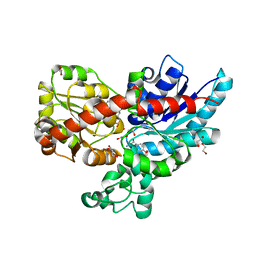
Crystal structure of UGT85B1 from Sorghum bicolor in complex with UDP and p-hydroxymandelonitrile
Descriptor: (2S)-HYDROXY(4-HYDROXYPHENYL)ETHANENITRILE, 1,2-ETHANEDIOL, Cyanohydrin beta-glucosyltransferase, ...
Authors:Putkaradze, N, Fredslund, F, Welner, D.H.
Deposit date:2022-03-31
Release date:2022-07-13
Last modified:2024-01-31
Method:X-RAY DIFFRACTION (1.5 Å)
Cite:Structure-guided engineering of key amino acids in UGT85B1 controlling substrate and stereo-specificity in aromatic cyanogenic glucoside biosynthesis.
Plant J., 111, 2022
8A58
DownloadVisualize
BU of 8a58 by Molmil
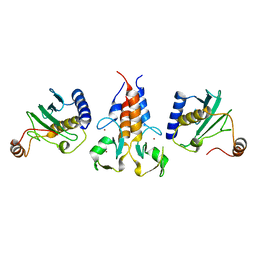
X-ray structure of TRIM21 RING E3 ligase in complex with E2 enzyme Ube2W
Descriptor: E3 ubiquitin-protein ligase TRIM21, Ubiquitin-conjugating enzyme E2 W, ZINC ION
Authors:James, L.C, Kiss, L.
Deposit date:2022-06-14
Release date:2023-04-26
Last modified:2024-02-07
Method:X-RAY DIFFRACTION (2.25 Å)
Cite:Trim-Away ubiquitinates and degrades lysine-less and N-terminally acetylated substrates.
Nat Commun, 14, 2023
7ZIE
DownloadVisualize
BU of 7zie by Molmil
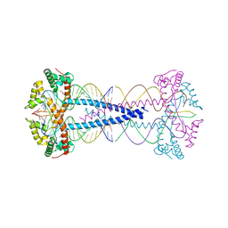
Gcf1p, multimerizes and bridges the mitochondrial DNA from Candida albicans by a specific mechanism.
Descriptor: 20-mer DNA, GLYCEROL, Gcf1p
Authors:Tarres-Sole, A, Sola, M.
Deposit date:2022-04-07
Release date:2023-06-14
Last modified:2024-06-19
Method:SOLUTION SCATTERING (2.9 Å), X-RAY DIFFRACTION
Cite:Gcf1p tetramerizes upon DNA binding, revealing a novel compaction method for mitochondrial DNA in Candida albicans.
Nucleic Acids Res., 2023
8A6W
DownloadVisualize
BU of 8a6w by Molmil
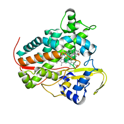
Crystal structure of CYP142 from Mycobacterium tuberculosis in complex with cholestenone
Descriptor: (8ALPHA,9BETA)-CHOLEST-4-EN-3-ONE, PROTOPORPHYRIN IX CONTAINING FE, Steroid C26-monooxygenase
Authors:Snee, M, Amadi, C, Levy, C.
Deposit date:2022-06-20
Release date:2023-07-05
Last modified:2024-02-07
Method:X-RAY DIFFRACTION (2.09 Å)
Cite:Crystal structure of CYP142 from Mycobacterium tuberculosis in complex with cholestenone
To Be Published
7ZJ5
DownloadVisualize
BU of 7zj5 by Molmil
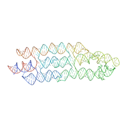
Unbound state of a brocolli-pepper aptamer FRET tile.
Descriptor: POTASSIUM ION, brocolli-pepper aptamer
Authors:McRae, E.K.S, Vallina, N.S, Hansen, B.K, Boussebayle, A, Andersen, E.S.
Deposit date:2022-04-08
Release date:2023-04-19
Last modified:2024-07-24
Method:ELECTRON MICROSCOPY (4.55 Å)
Cite:Structure determination of Pepper-Broccoli FRET pair by RNA origami scaffolding
To Be Published
8A67
DownloadVisualize
BU of 8a67 by Molmil
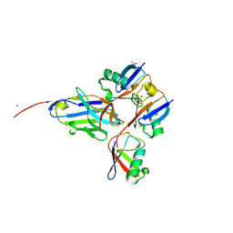
Branched Lys48- and Lys63-linked tri-ubiquitin (K48-K63-Ub3) in complex with matured synthetic nanobody NbSL3.3Q (3rd generation)
Descriptor: CHLORIDE ION, GLYCEROL, ISOPROPYL ALCOHOL, ...
Authors:Lange, S.M, Kulathu, Y.
Deposit date:2022-06-16
Release date:2023-02-15
Last modified:2024-10-16
Method:X-RAY DIFFRACTION (1.86 Å)
Cite:VCP/p97-associated proteins are binders and debranching enzymes of K48-K63-branched ubiquitin chains.
Nat.Struct.Mol.Biol., 2024

226707

PDB entries from 2024-10-30

PDB statisticsPDBj update infoContact PDBjnumon