Loading
PDBj
MenuPDBj@FacebookPDBj@TwitterPDBj@YouTubewwPDB FoundationwwPDB
RCSB PDBPDBeBMRBAdv. SearchSearch help
Search by PDB author
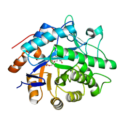
3EKD
DownloadVisualize
BU of 3ekd by Molmil
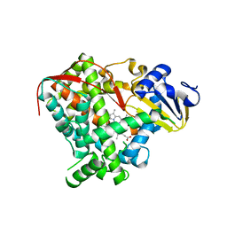
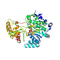
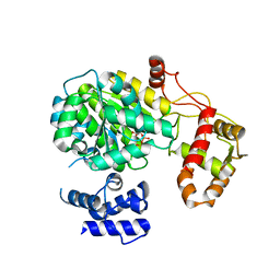
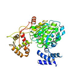
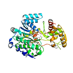
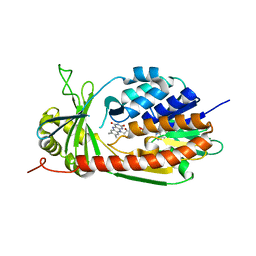
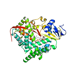
Crystal structure of the A264M heme domain of cytochrome P450 BM3
Descriptor: Cytochrome P450(BM-3), PALMITOLEIC ACID, PROTOPORPHYRIN IX CONTAINING FE
Authors:Toogood, H.S, Leys, D.
Deposit date:2008-09-19
Release date:2008-12-30
Last modified:2023-08-30
Method:X-RAY DIFFRACTION (2.5 Å)
Cite:Novel haem co-ordination variants of flavocytochrome P450BM3.
Biochem.J., 417, 2009
3F03
DownloadVisualize
BU of 3f03 by Molmil
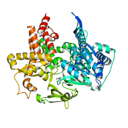
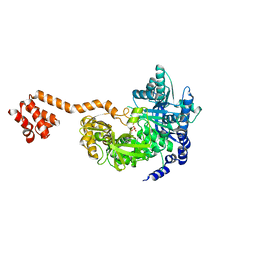
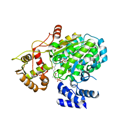
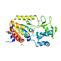
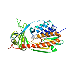
Crystal structure of Pentaerythritol Tetranitrate Reductase complex with 1-nitrocyclohexene
Descriptor: 1-nitrocyclohexene, CHLORIDE ION, FLAVIN MONONUCLEOTIDE, ...
Authors:Roujeinikova, A.R, Toogood, H.S, Leys, D.
Deposit date:2008-10-24
Release date:2008-12-16
Last modified:2023-12-27
Method:X-RAY DIFFRACTION (1.34 Å)
Cite:Structure-based insight into the asymmetric bioreduction of the C=C double bond of alpha,beta-unsaturated nitroalkenes by pentaerythritol tetranitrate reductase.
To be published
3EKF
DownloadVisualize
BU of 3ekf by Molmil
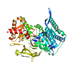
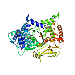
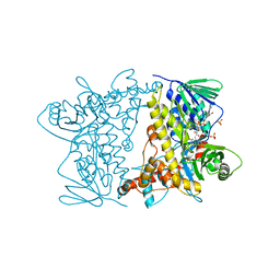
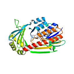
Crystal structure of the A264Q heme domain of cytochrome P450 BM3
Descriptor: Cytochrome P450(BM-3), PROTOPORPHYRIN IX CONTAINING FE
Authors:Toogood, H.S, Leys, D.
Deposit date:2008-09-19
Release date:2008-12-30
Last modified:2023-08-30
Method:X-RAY DIFFRACTION (2.1 Å)
Cite:Novel haem co-ordination variants of flavocytochrome P450BM3.
Biochem.J., 417, 2009
5MSS
DownloadVisualize
BU of 5mss by Molmil
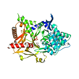
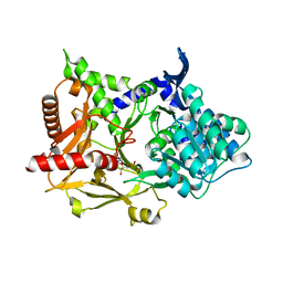
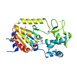
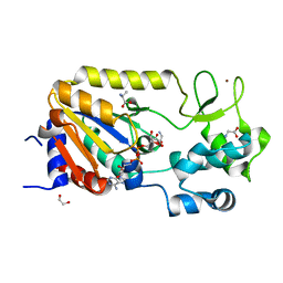
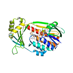
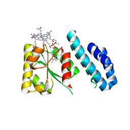
Structure of the A-PCP didomain of carboxylic acid reductase (CAR) from Segniliparus rugosus in complex with AMP
Descriptor: ADENOSINE MONOPHOSPHATE, SODIUM ION, Thioester reductase domain-containing protein
Authors:Gahloth, D, Leys, D.
Deposit date:2017-01-05
Release date:2017-07-05
Last modified:2024-01-17
Method:X-RAY DIFFRACTION (1.96 Å)
Cite:Structures of carboxylic acid reductase reveal domain dynamics underlying catalysis.
Nat. Chem. Biol., 13, 2017
5MST
DownloadVisualize
BU of 5mst by Molmil
Structure of the A domain of carboxylic acid reductase (CAR) from Segniliparus rugosus in complex with AMP and a co-purified carboxylic acid
Descriptor: ADENOSINE MONOPHOSPHATE, CALCIUM ION, FUMARIC ACID, ...
Authors:Gahloth, D, Leys, D.
Deposit date:2017-01-05
Release date:2017-07-05
Last modified:2024-01-17
Method:X-RAY DIFFRACTION (1.72 Å)
Cite:Structures of carboxylic acid reductase reveal domain dynamics underlying catalysis.
Nat. Chem. Biol., 13, 2017
5MSD
DownloadVisualize
BU of 5msd by Molmil
Structure of the A domain of carboxylic acid reductase (CAR) from Nocardia iowensis in complex with AMP and benzoic acid
Descriptor: ADENOSINE MONOPHOSPHATE, BENZOIC ACID, Carboxylic acid reductase
Authors:Dunstan, M.S, Leys, D.
Deposit date:2017-01-04
Release date:2017-07-05
Last modified:2024-01-17
Method:X-RAY DIFFRACTION (1.71 Å)
Cite:Structures of carboxylic acid reductase reveal domain dynamics underlying catalysis.
Nat. Chem. Biol., 13, 2017
5MSO
DownloadVisualize
BU of 5mso by Molmil
Structure of the R domain of carboxylic acid reductase (CAR) from Mycobacterium marinum in complex with NADP
Descriptor: Carboxylic acid reductase, NADP NICOTINAMIDE-ADENINE-DINUCLEOTIDE PHOSPHATE
Authors:Gahloth, D, Leys, D.
Deposit date:2017-01-05
Release date:2017-07-05
Last modified:2024-01-17
Method:X-RAY DIFFRACTION (1.2 Å)
Cite:Structures of carboxylic acid reductase reveal domain dynamics underlying catalysis.
Nat. Chem. Biol., 13, 2017
5MSW
DownloadVisualize
BU of 5msw by Molmil
Structure of the A-PCP didomain of carboxylic acid reductase (CAR) from Segniliparus rugosus in complex with AMP
Descriptor: ADENOSINE MONOPHOSPHATE, Thioester reductase domain-containing protein
Authors:Gahloth, D, Leys, D.
Deposit date:2017-01-05
Release date:2017-07-05
Last modified:2024-01-17
Method:X-RAY DIFFRACTION (2.33 Å)
Cite:Structures of carboxylic acid reductase reveal domain dynamics underlying catalysis.
Nat. Chem. Biol., 13, 2017
5MSQ
DownloadVisualize
BU of 5msq by Molmil
Structure of the A domain of carboxylic acid reductase (CAR) from Nocardia iowensis in complex with AMP and iodide
Descriptor: ADENOSINE MONOPHOSPHATE, Carboxylic acid reductase, IODIDE ION
Authors:Dunstan, M.S, Leys, D.
Deposit date:2017-01-05
Release date:2018-01-17
Last modified:2024-05-08
Method:X-RAY DIFFRACTION (1.74 Å)
Cite:Structure and mechanism of carboxylic acid reductase
To be published
5MSC
DownloadVisualize
BU of 5msc by Molmil
Structure of the A domain of carboxylic acid reductase (CAR) from Nocardia iowensis in complex with AMP
Descriptor: ADENOSINE MONOPHOSPHATE, Carboxylic acid reductase
Authors:Dunstan, M.S, Leys, D.
Deposit date:2017-01-04
Release date:2017-07-05
Last modified:2024-05-08
Method:X-RAY DIFFRACTION (1.85 Å)
Cite:Structures of carboxylic acid reductase reveal domain dynamics underlying catalysis.
Nat. Chem. Biol., 13, 2017
5MSP
DownloadVisualize
BU of 5msp by Molmil
Structure of the unmodified PCP-R didomain of carboxylic acid reductase (CAR) from Segniliparus rugosus in complex with NADP, F2221 form
Descriptor: NADP NICOTINAMIDE-ADENINE-DINUCLEOTIDE PHOSPHATE, Thioester reductase domain-containing protein
Authors:Gahloth, D, Leys, D.
Deposit date:2017-01-05
Release date:2017-07-05
Last modified:2024-01-17
Method:X-RAY DIFFRACTION (2.41 Å)
Cite:Structures of carboxylic acid reductase reveal domain dynamics underlying catalysis.
Nat. Chem. Biol., 13, 2017
5MSV
DownloadVisualize
BU of 5msv by Molmil
Structure of the phosphopantetheine modified PCP-R didomain of carboxylic acid reductase (CAR) in complex with NADP
Descriptor: 4'-PHOSPHOPANTETHEINE, NADP NICOTINAMIDE-ADENINE-DINUCLEOTIDE PHOSPHATE, Thioester reductase domain-containing protein
Authors:Gahloth, D, Leys, D.
Deposit date:2017-01-05
Release date:2017-07-05
Last modified:2024-01-17
Method:X-RAY DIFFRACTION (2.34 Å)
Cite:Structures of carboxylic acid reductase reveal domain dynamics underlying catalysis.
Nat. Chem. Biol., 13, 2017
4Z44
DownloadVisualize
BU of 4z44 by Molmil
F454K Mutant of Tryptophan 7-halogenase PrnA
Descriptor: CHLORIDE ION, FLAVIN-ADENINE DINUCLEOTIDE, Flavin-dependent tryptophan halogenase PrnA, ...
Authors:Shepherd, S.A, Karthikeyan, C, Latham, J, Struck, A.-W, Thompson, M.L, Menon, B, Levy, C.W, Leys, D, Micklefield, J.
Deposit date:2015-04-01
Release date:2016-04-13
Last modified:2024-01-10
Method:X-RAY DIFFRACTION (2.204 Å)
Cite:Extending the biocatalytic scope of regiocomplementary flavin-dependent halogenase enzymes.
Chem Sci, 6, 2015
5A3C
DownloadVisualize
BU of 5a3c by Molmil
Crystal structure of the ADP-ribosylating sirtuin (SirTM) from Streptococcus pyogenes in complex with NAD
Descriptor: 1,2-ETHANEDIOL, GLYCINE, NICOTINAMIDE-ADENINE-DINUCLEOTIDE, ...
Authors:Rack, J.G.M, Morra, R, Barkauskaite, E, Kraehenbuehl, R, Ariza, A, Qu, Y, Ortmayer, M, Leidecker, O, Cameron, D.R, Matic, I, Peleg, A.Y, Leys, D, Traven, A, Ahel, I.
Deposit date:2015-05-28
Release date:2015-07-29
Last modified:2024-05-08
Method:X-RAY DIFFRACTION (2.03 Å)
Cite:Identification of a Class of Protein Adp-Ribosylating Sirtuins in Microbial Pathogens.
Mol.Cell, 59, 2015
5MSR
DownloadVisualize
BU of 5msr by Molmil
Structure of the unmodified PCP-R domain of carboxylic acid reductase (CAR) from Segniliparus rugosus in complex with NADPH, P43 form
Descriptor: NADP NICOTINAMIDE-ADENINE-DINUCLEOTIDE PHOSPHATE, Thioester reductase domain-containing protein
Authors:Gahloth, D, Leys, D.
Deposit date:2017-01-05
Release date:2017-07-05
Last modified:2024-01-17
Method:X-RAY DIFFRACTION (2.37 Å)
Cite:Structures of carboxylic acid reductase reveal domain dynamics underlying catalysis.
Nat. Chem. Biol., 13, 2017
5A3A
DownloadVisualize
BU of 5a3a by Molmil
Crystal structure of the ADP-ribosylating sirtuin (SirTM) from Streptococcus pyogenes (Apo form)
Descriptor: 1,2-ETHANEDIOL, GLYCINE, SIR2 FAMILY PROTEIN, ...
Authors:Rack, J.G.M, Morra, R, Barkauskaite, E, Kraehenbuehl, R, Ariza, A, Qu, Y, Ortmayer, M, Leidecker, O, Cameron, D.R, Matic, I, Peleg, A.Y, Leys, D, Traven, A, Ahel, I.
Deposit date:2015-05-28
Release date:2015-07-29
Last modified:2024-05-08
Method:X-RAY DIFFRACTION (1.54 Å)
Cite:Identification of a Class of Protein Adp-Ribosylating Sirtuins in Microbial Pathogens.
Mol.Cell, 59, 2015
5A3B
DownloadVisualize
BU of 5a3b by Molmil
Crystal structure of the ADP-ribosylating sirtuin (SirTM) from Streptococcus pyogenes in complex with ADP-ribose
Descriptor: 1,2-ETHANEDIOL, ADENOSINE-5-DIPHOSPHORIBOSE, ALANINE, ...
Authors:Rack, J.G.M, Morra, R, Barkauskaite, E, Kraehenbuehl, R, Ariza, A, Qu, Y, Ortmayer, M, Leidecker, O, Cameron, D.R, Matic, I, Peleg, A.Y, Leys, D, Traven, A, Ahel, I.
Deposit date:2015-05-28
Release date:2015-07-29
Last modified:2024-05-08
Method:X-RAY DIFFRACTION (1.9 Å)
Cite:Identification of a Class of Protein Adp-Ribosylating Sirtuins in Microbial Pathogens.
Mol.Cell, 59, 2015
5A35
DownloadVisualize
BU of 5a35 by Molmil
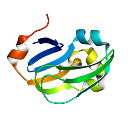
Crystal structure of Glycine Cleavage Protein H-Like (GcvH-L) from Streptococcus pyogenes
Descriptor: GLYCINE CLEAVAGE SYSTEM H PROTEIN, PENTAETHYLENE GLYCOL
Authors:Rack, J.G.M, Morra, R, Barkauskaite, E, Kraehenbuehl, R, Ariza, A, Qu, Y, Ortmayer, M, Leidecker, O, Cameron, D.R, Matic, I, Peleg, A.Y, Leys, D, Traven, A, Ahel, I.
Deposit date:2015-05-27
Release date:2015-07-29
Last modified:2024-01-10
Method:X-RAY DIFFRACTION (1.5 Å)
Cite:Identification of a Class of Protein Adp-Ribosylating Sirtuins in Microbial Pathogens.
Mol.Cell, 59, 2015
5MSU
DownloadVisualize
BU of 5msu by Molmil
Structure of the R domain of carboxylic acid reductase (CAR) from Mycobacterium marinum in complex with NADP, P21 form
Descriptor: Carboxylic acid reductase, NADP NICOTINAMIDE-ADENINE-DINUCLEOTIDE PHOSPHATE
Authors:Gahloth, D, Leys, D.
Deposit date:2017-01-05
Release date:2017-07-05
Last modified:2024-01-17
Method:X-RAY DIFFRACTION (1.74 Å)
Cite:Structures of carboxylic acid reductase reveal domain dynamics underlying catalysis.
Nat. Chem. Biol., 13, 2017
4J2W
DownloadVisualize
BU of 4j2w by Molmil
Crystal Structure of kynurenine 3-monooxygenase (KMO-396Prot-Se)
Descriptor: FLAVIN-ADENINE DINUCLEOTIDE, Kynurenine 3-monooxygenase
Authors:Amaral, M, Levy, C, Heyes, D.J, Lafite, P, Outeiro, T.F, Giorgini, F, Leys, D, Scrutton, N.S.
Deposit date:2013-02-05
Release date:2013-04-10
Last modified:2024-02-28
Method:X-RAY DIFFRACTION (2.6 Å)
Cite:Structural basis of kynurenine 3-monooxygenase inhibition.
Nature, 496, 2013
4J31
DownloadVisualize
BU of 4j31 by Molmil
Crystal Structure of kynurenine 3-monooxygenase (KMO-396Prot)
Descriptor: FLAVIN-ADENINE DINUCLEOTIDE, Kynurenine 3-monooxygenase
Authors:Amaral, M, Levy, C, Heyes, D.J, Lafite, P, Outeiro, T.F, Giorgini, F, Leys, D, Scrutton, N.S.
Deposit date:2013-02-05
Release date:2013-04-10
Last modified:2024-05-22
Method:X-RAY DIFFRACTION (2.4 Å)
Cite:Structural basis of kynurenine 3-monooxygenase inhibition.
Nature, 496, 2013
4J36
DownloadVisualize
BU of 4j36 by Molmil
Cocrystal Structure of kynurenine 3-monooxygenase in complex with UPF 648 inhibitor(KMO-394UPF)
Descriptor: (1S,2S)-2-(3,4-dichlorobenzoyl)cyclopropanecarboxylic acid, FLAVIN-ADENINE DINUCLEOTIDE, Kynurenine 3-monooxygenase
Authors:Amaral, M, Levy, C, Heyes, D.J, Lafite, P, Outeiro, T.F, Giorgini, F, Leys, D, Scrutton, N.S.
Deposit date:2013-02-05
Release date:2013-04-10
Last modified:2024-02-28
Method:X-RAY DIFFRACTION (2.13 Å)
Cite:Structural basis of kynurenine 3-monooxygenase inhibition.
Nature, 496, 2013
4J33
DownloadVisualize
BU of 4j33 by Molmil
Crystal Structure of kynurenine 3-monooxygenase (KMO-394)
Descriptor: FLAVIN-ADENINE DINUCLEOTIDE, Kynurenine 3-monooxygenase
Authors:Amaral, M, Levy, C, Heyes, D.J, Lafite, P, Outeiro, T.F, Giorgini, F, Leys, D, Scrutton, N.S.
Deposit date:2013-02-05
Release date:2013-04-10
Last modified:2024-02-28
Method:X-RAY DIFFRACTION (1.82 Å)
Cite:Structural basis of kynurenine 3-monooxygenase inhibition.
Nature, 496, 2013
4JGI
DownloadVisualize
BU of 4jgi by Molmil
1.5 Angstrom crystal structure of a novel cobalamin-binding protein from Desulfitobacterium hafniense DCB-2
Descriptor: CO-METHYLCOBALAMIN, Putative uncharacterized protein
Authors:Sjuts, H, Dunstan, M.S, Fisher, K, Leys, D.
Deposit date:2013-03-01
Release date:2013-08-07
Last modified:2024-02-28
Method:X-RAY DIFFRACTION (1.5 Å)
Cite:Structure of the cobalamin-binding protein of a putative O-demethylase from Desulfitobacterium hafniense DCB-2.
Acta Crystallogr.,Sect.D, 69, 2013
4LX6
DownloadVisualize
BU of 4lx6 by Molmil
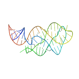
X-ray crystal structure of the M6C" riboswitch aptamer bound to 2-aminopyrimido[4,5-d]pyrimidin-4(3H)-one (PPAO)
Descriptor: 2-aminopyrimido[4,5-d]pyrimidin-4(3H)-one, MAGNESIUM ION, Mutated adenine riboswitch aptamer
Authors:Dunstan, M.S, Leys, D.
Deposit date:2013-07-29
Release date:2014-07-16
Last modified:2023-09-20
Method:X-RAY DIFFRACTION (2.15 Å)
Cite:Modular riboswitch toolsets for synthetic genetic control in diverse bacterial species.
J.Am.Chem.Soc., 136, 2014

226707

PDB entries from 2024-10-30

PDB statisticsPDBj update infoContact PDBjnumon