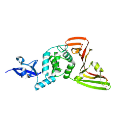
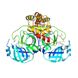
9RHO
 
 | | Structure of SARS-coV-2 NSP3 macrodomain in complex with ligand | | Descriptor: | Papain-like protease nsp3, [[(2~{R},3~{S},4~{R},5~{R})-5-(4-azanylpyrrolo[2,1-f][1,2,4]triazin-7-yl)-5-cyano-3,4-bis(oxidanyl)oxolan-2-yl]methoxy-oxidanyl-phosphoryl] methyl hydrogen phosphate | | Authors: | Ruiz Carrillo, D, Sander, S, Tidow, H, Garcia-Alai, M. | | Deposit date: | 2025-06-09 | | Release date: | 2026-06-24 | | Method: | X-RAY DIFFRACTION (1.6 Å) | | Cite: | Structure of SARS-coV-2 NSP3 macrodomain in complex with ligand
To Be Published
|
|
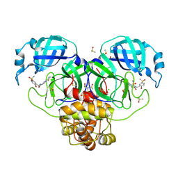
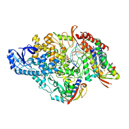
9VHT
 | | The crystal structure of PLPro | | Descriptor: | CHLORIDE ION, MALONATE ION, Papain-like protease nsp3, ... | | Authors: | Xiao, Q.J. | | Deposit date: | 2025-06-17 | | Release date: | 2026-06-24 | | Method: | X-RAY DIFFRACTION (1.82 Å) | | Cite: | The crystal structure of PLPro
To Be Published
|
|
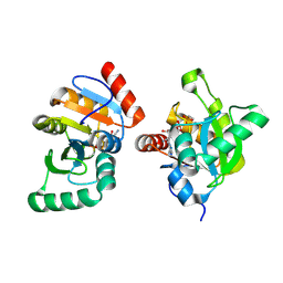
29OK
 | | PanDDA deposition -- Crystal Structure of SARS-CoV-2 main protease covalently bound to KL_C172 | | Descriptor: | 3C-like proteinase nsp5, CHLORIDE ION, DIMETHYL SULFOXIDE, ... | | Authors: | Balcomb, B.H, Aschenbrenner, J.C, Kollar, L, Mihalovits, L.M, Tomlinson, C.W.E, Marples, P.G, Fairhead, M, Fearon, D, von delft, F, Bajusz, D, Keseru, G.M. | | Deposit date: | 2026-03-26 | | Release date: | 2026-06-24 | | Method: | X-RAY DIFFRACTION (1.93 Å) | | Cite: | Fragment-Based Design of Targeted Covalent Inhibitors: The Scope and Limitation of Linking Approaches.
Chemmedchem, 21, 2026
|
|
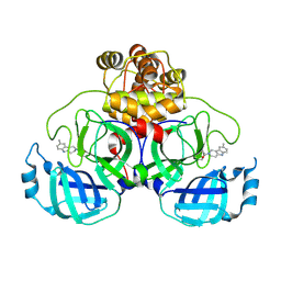
9RHN
 | | Structure of SARS-coV-2 NSP3 macrodomain in complex with ligand | | Descriptor: | Papain-like protease nsp3, [[(2~{R},3~{S},4~{R},5~{R})-5-(4-azanylpyrrolo[2,1-f][1,2,4]triazin-7-yl)-5-cyano-3,4-bis(oxidanyl)oxolan-2-yl]methoxy-oxidanyl-phosphoryl]oxy-ethyl-phosphinic acid | | Authors: | Ruiz Carrillo, D, Sander, S, Tidow, H, Garcia-Alai, M, Fliegert, R, Sandmann, M. | | Deposit date: | 2025-06-09 | | Release date: | 2026-06-24 | | Method: | X-RAY DIFFRACTION (1.95 Å) | | Cite: | Structure of SARS-coV-2 NSP3 macrodomain in complex with ligand
To Be Published
|
|
9OT4
 | | SARS-CoV-2 Main Protease (Mpro) in Complex with Covalent Inhibitor E9 | | Descriptor: | 1-(8-methyl-1,3,4,5-tetrahydro-2H-pyrido[4,3-b]indol-2-yl)ethan-1-one, 3C-like proteinase nsp5 | | Authors: | Mazzorana, M, Oliveira Borges, P.H, Silva-Junior, F, Baptista Ferreira, S, Walsh, M.A. | | Deposit date: | 2025-05-26 | | Release date: | 2026-06-24 | | Method: | X-RAY DIFFRACTION (2.1 Å) | | Cite: | SARS-CoV-2 Main Protease (Mpro) in Complex with Covalent Inhibitor E2
To Be Published
|
|
9OT5
 | | SARS-CoV-2 Main Protease (Mpro) in Complex with Covalent Inhibitor E2 | | Descriptor: | 1-[1-(4-chlorophenyl)-2,5-dimethyl-1H-pyrrol-3-yl]ethan-1-one, 3C-like proteinase nsp5, DI(HYDROXYETHYL)ETHER, ... | | Authors: | Mazzorana, M, Oliveira Borges, P.H, Silva-Junior, F, Baptista Ferreira, S, Walsh, M.A. | | Deposit date: | 2025-05-26 | | Release date: | 2026-06-24 | | Method: | X-RAY DIFFRACTION (2.5 Å) | | Cite: | SARS-CoV-2 Main Protease (Mpro) in Complex with Covalent Inhibitor E2
To Be Published
|
|
12SN
 | |


