 Movie
Movie Controller


+ Open data

- Basic information
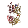
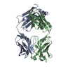
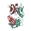
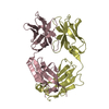
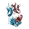
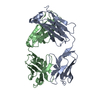
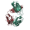
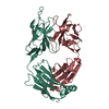
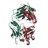
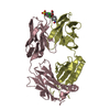
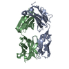
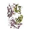
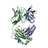
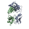
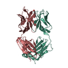
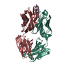
| Entry | Database: PDB / ID: 6pe7 | ||||||
|---|---|---|---|---|---|---|---|
| Title | Crystal Structure of ABBV-323 FAB | ||||||
 Components Components |
| ||||||
 Keywords Keywords | IMMUNE SYSTEM / CD40 / FAB | ||||||
| Function / homology | Immunoglobulins / Immunoglobulin-like / Sandwich / Mainly Beta Function and homology information Function and homology information | ||||||
| Biological species |  Homo sapiens (human) Homo sapiens (human) | ||||||
| Method |  X-RAY DIFFRACTION / SYNCHROTRON / MOLECULAR REPLACEMENT / Resolution: 1.74 Å X-RAY DIFFRACTION / SYNCHROTRON / MOLECULAR REPLACEMENT / Resolution: 1.74 Å | ||||||
 Authors Authors | Argiriadi, M.A. | ||||||
 Citation Citation | Journal: BMC Mol Cell Biol / Year: 2019 Title: CD40/anti-CD40 antibody complexes which illustrate agonist and antagonist structural switches. Authors: Argiriadi, M.A. / Benatuil, L. / Dubrovska, I. / Egan, D.A. / Gao, L. / Greischar, A. / Hardman, J. / Harlan, J. / Iyer, R.B. / Judge, R.A. / Lake, M. / Perron, D.C. / Sadhukhan, R. / ...Authors: Argiriadi, M.A. / Benatuil, L. / Dubrovska, I. / Egan, D.A. / Gao, L. / Greischar, A. / Hardman, J. / Harlan, J. / Iyer, R.B. / Judge, R.A. / Lake, M. / Perron, D.C. / Sadhukhan, R. / Sielaff, B. / Sousa, S. / Wang, R. / McRae, B.L. | ||||||
| History |
|
- Structure visualization
| Structure viewer | Molecule: MolmilJmol/JSmol |
|---|
- Downloads & links
-Download
| PDBx/mmCIF format | 6pe7.cif.gz | 106.1 KB | Display | PDBx/mmCIF format |
|---|---|---|---|---|
| PDB format | pdb6pe7.ent.gz | 78.7 KB | Display | PDB format |
| PDBx/mmJSON format | 6pe7.json.gz | Tree view | PDBx/mmJSON format | |
| Others |  Other downloads Other downloads |
-Validation report
| Arichive directory | https://data.pdbj.org/pub/pdb/validation_reports/pe/6pe7ftp://data.pdbj.org/pub/pdb/validation_reports/pe/6pe7 | HTTPS FTP |
|---|
-Related structure data
| Related structure data |  6pe8C  6pe9C  3qos S: Starting model for refinement C: citing same article ( |
|---|---|
| Similar structure data |


-Links
 PDBj
PDBj

- Assembly
| Deposited unit | 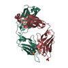
| ||||||||
|---|---|---|---|---|---|---|---|---|---|
| 1 |
| ||||||||
| Unit cell |
|
-Components
| #1: Antibody | Mass: 23748.588 Da / Num. of mol.: 1 Source method: isolated from a genetically manipulated source Source: (gene. exp.) Homo sapiens (human) / Cell line (production host): HEK293 / Production host: Homo sapiens (human) | ||||||
|---|---|---|---|---|---|---|---|
| #2: Antibody | Mass: 24290.963 Da / Num. of mol.: 1 Source method: isolated from a genetically manipulated source Source: (gene. exp.) Homo sapiens (human) / Cell line (production host): HEK293 / Production host: Homo sapiens (human) | ||||||
| #3: Chemical |   Mass: 96.063 Da / Num. of mol.: 2 / Source method: obtained synthetically / Formula: SO4 Mass: 96.063 Da / Num. of mol.: 2 / Source method: obtained synthetically / Formula: SO4#4: Water | ChemComp-HOH / |  Mass: 18.015 Da / Num. of mol.: 394 / Source method: isolated from a natural source / Formula: H2O Mass: 18.015 Da / Num. of mol.: 394 / Source method: isolated from a natural source / Formula: H2OHas ligand of interest | N | Has protein modification | Y | |
-Experimental details
-Experiment
| Experiment | Method: X-RAY DIFFRACTION / Number of used crystals: 1 |
|---|
- Sample preparation
| Crystal | Density Matthews: 2.86 Å3/Da / Density % sol: 57.06 % |
|---|---|
| Crystal grow | Temperature: 296.15 K / Method: vapor diffusion Details: 25% (w/v) PMME 550, 0.1M MES pH 6.5, 0.01M zinc sulfate |
-Data collection
| Diffraction | Mean temperature: 100 K / Serial crystal experiment: N |
|---|---|
| Diffraction source | Source: SYNCHROTRON / Site: APS  / Beamline: 17-ID / Wavelength: 1 Å / Beamline: 17-ID / Wavelength: 1 Å |
| Detector | Type: DECTRIS PILATUS 6M / Detector: PIXEL / Date: Dec 4, 2014 |
| Radiation | Protocol: SINGLE WAVELENGTH / Monochromatic (M) / Laue (L): M / Scattering type: x-ray |
| Radiation wavelength | Wavelength: 1 Å / Relative weight: 1 |
| Reflection | Resolution: 1.74→57.2 Å / Num. obs: 56956 / % possible obs: 100 % / Redundancy: 6.6 % / CC1/2: 1 / Net I/σ(I): 25.5 |
| Reflection shell | Resolution: 1.74→1.75 Å / Num. unique obs: 545 / CC1/2: 0.793 |
- Processing
| Software |
| ||||||||||||||||||||||||||||||||||||||||||||||||||||||||||||||||||||||||||||||||||||||||||||||||||||||||||||||||||||||||||||||||||||||||||||
|---|---|---|---|---|---|---|---|---|---|---|---|---|---|---|---|---|---|---|---|---|---|---|---|---|---|---|---|---|---|---|---|---|---|---|---|---|---|---|---|---|---|---|---|---|---|---|---|---|---|---|---|---|---|---|---|---|---|---|---|---|---|---|---|---|---|---|---|---|---|---|---|---|---|---|---|---|---|---|---|---|---|---|---|---|---|---|---|---|---|---|---|---|---|---|---|---|---|---|---|---|---|---|---|---|---|---|---|---|---|---|---|---|---|---|---|---|---|---|---|---|---|---|---|---|---|---|---|---|---|---|---|---|---|---|---|---|---|---|---|---|---|
| Refinement | Method to determine structure: MOLECULAR REPLACEMENT Starting model: 3QOS 3qos Resolution: 1.74→24.356 Å / SU ML: 0.22 / Cross valid method: FREE R-VALUE / σ(F): 1.9 / Phase error: 22.71 / Stereochemistry target values: MLHL
| ||||||||||||||||||||||||||||||||||||||||||||||||||||||||||||||||||||||||||||||||||||||||||||||||||||||||||||||||||||||||||||||||||||||||||||
| Solvent computation | Shrinkage radii: 0.9 Å / VDW probe radii: 1.11 Å / Solvent model: FLAT BULK SOLVENT MODEL | ||||||||||||||||||||||||||||||||||||||||||||||||||||||||||||||||||||||||||||||||||||||||||||||||||||||||||||||||||||||||||||||||||||||||||||
| Refinement step | Cycle: LAST / Resolution: 1.74→24.356 Å
| ||||||||||||||||||||||||||||||||||||||||||||||||||||||||||||||||||||||||||||||||||||||||||||||||||||||||||||||||||||||||||||||||||||||||||||
| Refine LS restraints |
| ||||||||||||||||||||||||||||||||||||||||||||||||||||||||||||||||||||||||||||||||||||||||||||||||||||||||||||||||||||||||||||||||||||||||||||
| LS refinement shell |
|
