 Movie
Movie Controller


+ Open data

- Basic information
| Entry | Database: PDB / ID: 6kdi | ||||||
|---|---|---|---|---|---|---|---|
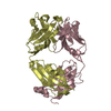
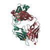
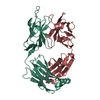
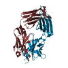
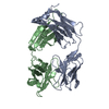
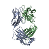
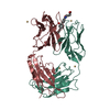
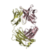
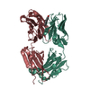
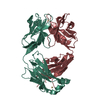
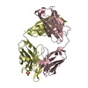
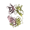
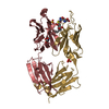
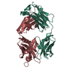
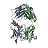
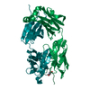
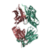
| Title | Antibody 64M-5 Fab including isoAsp in complex with dT(6-4)T | ||||||
 Components Components |
| ||||||
 Keywords Keywords | IMMUNE SYSTEM / DNA (6-4) PHOTOPRODUCT / IMMUNOGLOBULIN / FAB / isoaspartate | ||||||
| Function / homology | Immunoglobulins / Immunoglobulin-like / Sandwich / Mainly Beta / DNA Function and homology information Function and homology information | ||||||
| Biological species |  synthetic construct (others) | ||||||
| Method |  X-RAY DIFFRACTION / MOLECULAR REPLACEMENT / Resolution: 2.7 Å X-RAY DIFFRACTION / MOLECULAR REPLACEMENT / Resolution: 2.7 Å | ||||||
 Authors Authors | Yokoyama, H. / Mizutani, R. / Noguchi, S. / Hayashida, N. | ||||||
| Funding support |  Japan, 1items Japan, 1items
| ||||||
 Citation Citation | Journal: Sci Rep / Year: 2019 Title: Structural and biochemical basis of the formation of isoaspartate in the complementarity-determining region of antibody 64M-5 Fab. Authors: Yokoyama, H. / Mizutani, R. / Noguchi, S. / Hayashida, N. #1: Journal: Acta Crystallogr F Struct Biol Commun / Year: 2019Title: Structures of the antibody 64M-5 Fab and its complex with dT(6-4)T indicate induced-fit and high-affinity mechanisms. Authors: Yokoyama, H. / Mizutani, R. / Noguchi, S. / Hayashida, N. | ||||||
| History |
|
- Structure visualization
| Structure viewer | Molecule: MolmilJmol/JSmol |
|---|
- Downloads & links
-Download
| PDBx/mmCIF format | 6kdi.cif.gz | 102.1 KB | Display | PDBx/mmCIF format |
|---|---|---|---|---|
| PDB format | pdb6kdi.ent.gz | 74.3 KB | Display | PDB format |
| PDBx/mmJSON format | 6kdi.json.gz | Tree view | PDBx/mmJSON format | |
| Others |  Other downloads Other downloads |
-Validation report
| Arichive directory | https://data.pdbj.org/pub/pdb/validation_reports/kd/6kdiftp://data.pdbj.org/pub/pdb/validation_reports/kd/6kdi | HTTPS FTP |
|---|
-Related structure data
| Related structure data | 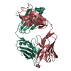 6kdhC 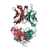 1ehlS S: Starting model for refinement C: citing same article ( |
|---|---|
| Similar structure data |


-Links
 PDBj
PDBj

- Assembly
| Deposited unit | 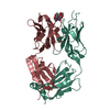
| ||||||||
|---|---|---|---|---|---|---|---|---|---|
| 1 |
| ||||||||
| Unit cell |
|
-Components
| #1: Antibody | Mass: 24097.699 Da / Num. of mol.: 1 / Source method: isolated from a natural source / Source: (natural) |
|---|---|
| #2: Antibody | Mass: 23828.641 Da / Num. of mol.: 1 / Source method: isolated from a natural source / Source: (natural) |
| #3: DNA chain | Mass: 565.445 Da / Num. of mol.: 1 / Source method: obtained synthetically / Source: (synth.) synthetic construct (others) |
| #4: Water | ChemComp-HOH /  Mass: 18.015 Da / Num. of mol.: 93 / Source method: isolated from a natural source / Formula: H2O Mass: 18.015 Da / Num. of mol.: 93 / Source method: isolated from a natural source / Formula: H2O |
| Has ligand of interest | Y |
| Has protein modification | Y |
-Experimental details
-Experiment
| Experiment | Method: X-RAY DIFFRACTION / Number of used crystals: 1 |
|---|
- Sample preparation
| Crystal | Density Matthews: 2.41 Å3/Da / Density % sol: 49.06 % |
|---|---|
| Crystal grow | Temperature: 293 K / Method: vapor diffusion, sitting drop / pH: 3.5 Details: 15% PEG2000, 0.1 M ammonium sulfate, 0.1 M sodium citrate |
-Data collection
| Diffraction | Mean temperature: 105 K / Serial crystal experiment: N |
|---|---|
| Diffraction source | Source: ROTATING ANODE / Type: MACSCIENCE / Wavelength: 1.5418 Å |
| Detector | Type: RIGAKU RAXIS IV / Detector: IMAGE PLATE / Date: Sep 22, 2000 |
| Radiation | Monochromator: GRAPHITE / Protocol: SINGLE WAVELENGTH / Monochromatic (M) / Laue (L): M / Scattering type: x-ray |
| Radiation wavelength | Wavelength: 1.5418 Å / Relative weight: 1 |
| Reflection | Resolution: 2.7→30 Å / Num. obs: 12638 / % possible obs: 95.2 % / Redundancy: 3.7 % / Rmerge(I) obs: 0.09 / Net I/σ(I): 14.5 |
| Reflection shell | Resolution: 2.7→2.8 Å / Rmerge(I) obs: 0.332 / Mean I/σ(I) obs: 2.9 / Num. unique obs: 967 / % possible all: 75 |
- Processing
| Software |
| ||||||||||||||||||||||||||||||||||||
|---|---|---|---|---|---|---|---|---|---|---|---|---|---|---|---|---|---|---|---|---|---|---|---|---|---|---|---|---|---|---|---|---|---|---|---|---|---|
| Refinement | Method to determine structure: MOLECULAR REPLACEMENT Starting model: 1EHL Resolution: 2.7→27.29 Å / Rfactor Rfree error: 0.007 / Data cutoff high absF: 57157 / Data cutoff low absF: 0 / Cross valid method: THROUGHOUT / σ(F): 0 / Details: BULK SOLVENT MODEL USED
| ||||||||||||||||||||||||||||||||||||
| Solvent computation | Bsol: 32.7839 Å2 / ksol: 0.35 e/Å3 | ||||||||||||||||||||||||||||||||||||
| Displacement parameters | Biso max: 76.4 Å2 / Biso mean: 37.9 Å2 / Biso min: 7.04 Å2
| ||||||||||||||||||||||||||||||||||||
| Refine analyze |
| ||||||||||||||||||||||||||||||||||||
| Refinement step | Cycle: final / Resolution: 2.7→27.29 Å
| ||||||||||||||||||||||||||||||||||||
| Refine LS restraints |
| ||||||||||||||||||||||||||||||||||||
| LS refinement shell | Resolution: 2.7→2.87 Å / Rfactor Rfree error: 0.03 / Total num. of bins used: 6
| ||||||||||||||||||||||||||||||||||||
| Xplor file |
|
