 Movie
Movie Controller

[English] 日本語
 Yorodumi
- PDB-1riv: Anti-Cocaine Antibody M82G2 Complexed With meta-Oxybenzoylecgonine -
+ Open data

- Basic information
| Entry | Database: PDB / ID: 1riv | ||||||
|---|---|---|---|---|---|---|---|
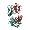
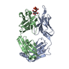
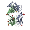
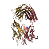
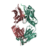
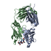
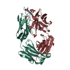
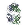
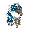
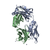
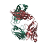
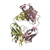
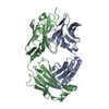
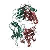
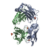
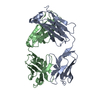
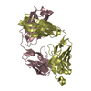
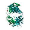
| Title | Anti-Cocaine Antibody M82G2 Complexed With meta-Oxybenzoylecgonine | ||||||
 Components Components |
| ||||||
 Keywords Keywords | IMMUNE SYSTEM / ANTI-COCAINE ANTIBODY / FAB | ||||||
| Function / homology | Immunoglobulins / Immunoglobulin-like / Sandwich / Mainly Beta / Chem-OBE Function and homology information Function and homology information | ||||||
| Biological species |  | ||||||
| Method |  X-RAY DIFFRACTION / MOLECULAR REPLACEMENT / Resolution: 2.2 Å X-RAY DIFFRACTION / MOLECULAR REPLACEMENT / Resolution: 2.2 Å | ||||||
 Authors Authors | Pozharski, E. / Hewagama, A. / Shanafelt, A. / Petsko, G. / Ringe, D. | ||||||
 Citation Citation | Journal: To be Published Title: Carving a Binding Site: Structural Study of an Anti-Cocaine Antibody in Complex with Three Cocaine Analogs Authors: Pozharski, E. / Hewagama, A. / Shanafelt, A. / Petsko, G. / Ringe, D. | ||||||
| History |
| ||||||
| Remark 999 | SEQUENCE A suitable database reference sequence could not be found for chains L and H at the time ...SEQUENCE A suitable database reference sequence could not be found for chains L and H at the time of processing. |
- Structure visualization
| Structure viewer | Molecule: MolmilJmol/JSmol |
|---|
- Downloads & links
-Download
| PDBx/mmCIF format | 1riv.cif.gz | 101.2 KB | Display | PDBx/mmCIF format |
|---|---|---|---|---|
| PDB format | pdb1riv.ent.gz | 76 KB | Display | PDB format |
| PDBx/mmJSON format | 1riv.json.gz | Tree view | PDBx/mmJSON format | |
| Others |  Other downloads Other downloads |
-Validation report
| Arichive directory | https://data.pdbj.org/pub/pdb/validation_reports/ri/1rivftp://data.pdbj.org/pub/pdb/validation_reports/ri/1riv | HTTPS FTP |
|---|
-Related structure data
| Related structure data | 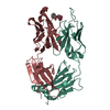 1riuC  1ar3  1axtS C: citing same article ( S: Starting model for refinement |
|---|---|
| Similar structure data |


-Links
 PDBj
PDBj

- Assembly
| Deposited unit | 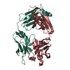
| ||||||||
|---|---|---|---|---|---|---|---|---|---|
| 1 |
| ||||||||
| Unit cell |
| ||||||||
| Components on special symmetry positions |
|
-Components
| #1: Antibody | Mass: 23884.365 Da / Num. of mol.: 1 / Source method: isolated from a natural source / Source: (natural) |
|---|---|
| #2: Antibody | Mass: 23519.264 Da / Num. of mol.: 1 / Source method: isolated from a natural source / Source: (natural) |
| #3: Chemical | ChemComp-OBE /   Mass: 305.326 Da / Num. of mol.: 1 / Source method: obtained synthetically / Formula: C16H19NO5 Mass: 305.326 Da / Num. of mol.: 1 / Source method: obtained synthetically / Formula: C16H19NO5 |
| #4: Water | ChemComp-HOH /  Mass: 18.015 Da / Num. of mol.: 182 / Source method: isolated from a natural source / Formula: H2O Mass: 18.015 Da / Num. of mol.: 182 / Source method: isolated from a natural source / Formula: H2O |
| Has protein modification | Y |
-Experimental details
-Experiment
| Experiment | Method: X-RAY DIFFRACTION / Number of used crystals: 1 |
|---|
- Sample preparation
| Crystal | Density Matthews: 2.57 Å3/Da / Density % sol: 51.7 % Description: CRYSTALS WERE TRANSFERRED STEPWISE INTO 70% SATURATED SODIUM MALONATE, PH 7.2, AND SUBSEQUENTLY SOAKED OVERNIGHT IN THE SODIUM MALONATE SOLUTION CONTAINING 300 MICROMOLAR OXYBENZOYLECGONINE-HCL |
|---|---|
| Crystal grow | Temperature: 298 K / Method: vapor diffusion, hanging drop / pH: 5.2 Details: 2M AMMONIUM SULFATE, 5% ISO-PROPANOL, pH 5.20, VAPOR DIFFUSION, HANGING DROP, temperature 298K |
-Data collection
| Diffraction | Mean temperature: 277 K |
|---|---|
| Diffraction source | Source: ROTATING ANODE / Type: RIGAKU RU300 / Wavelength: 1.5418 Å |
| Detector | Type: RIGAKU RAXIS IV / Detector: IMAGE PLATE / Date: Aug 20, 2002 / Details: mirrors |
| Radiation | Monochromator: graphite / Protocol: SINGLE WAVELENGTH / Monochromatic (M) / Laue (L): M / Scattering type: x-ray |
| Radiation wavelength | Wavelength: 1.5418 Å / Relative weight: 1 |
| Reflection | Resolution: 2.2→38 Å / Num. all: 26936 / Num. obs: 26936 / % possible obs: 99.8 % / Observed criterion σ(F): 0 / Observed criterion σ(I): 0 / Redundancy: 9.1 % / Biso Wilson estimate: 43.8 Å2 / Rsym value: 0.119 / Net I/σ(I): 13.7 |
| Reflection shell | Resolution: 2.2→2.28 Å / Redundancy: 4.62 % / Mean I/σ(I) obs: 1.97 / Num. unique all: 2637 / % possible all: 99.7 |
- Processing
| Software |
| ||||||||||||||||||||||||||||||||||||
|---|---|---|---|---|---|---|---|---|---|---|---|---|---|---|---|---|---|---|---|---|---|---|---|---|---|---|---|---|---|---|---|---|---|---|---|---|---|
| Refinement | Method to determine structure: MOLECULAR REPLACEMENT Starting model: PDB ENTRY 1AR3 (CONSTANT DOMAIN) + PDB ENTRY 1AXT (VARIABLE DOMAIN) Resolution: 2.2→38 Å / Isotropic thermal model: ISOTROPIC / Cross valid method: THROUGHOUT / σ(F): 0 / Stereochemistry target values: ENGH & HUBER Details: BULK SOLVENT MASK CORRECTED TO EXCLUDE INTERNAL CAVITIES
| ||||||||||||||||||||||||||||||||||||
| Solvent computation | Solvent model: MASK / Bsol: 48.9 Å2 / ksol: 0.36 e/Å3 | ||||||||||||||||||||||||||||||||||||
| Displacement parameters | Biso mean: 33.7 Å2
| ||||||||||||||||||||||||||||||||||||
| Refine analyze |
| ||||||||||||||||||||||||||||||||||||
| Refinement step | Cycle: LAST / Resolution: 2.2→38 Å
| ||||||||||||||||||||||||||||||||||||
| Refine LS restraints |
| ||||||||||||||||||||||||||||||||||||
| LS refinement shell | Resolution: 2.2→2.28 Å / Total num. of bins used: 10
|
