 Movie
Movie Controller


+ Open data

- Basic information
| Entry | Database: PDB / ID: 4e9o | ||||||
|---|---|---|---|---|---|---|---|
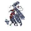
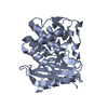
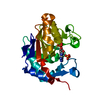
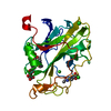
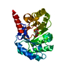
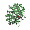
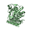
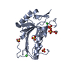
| Title | Vaccinia D8L ectodomain structure | ||||||
 Components Components | IMV membrane protein | ||||||
 Keywords Keywords | VIRAL PROTEIN / CAH alpha fold / Vp7 motif / beta sheet / cell surface chondroitin binding / viral entry / chondroitin sulfate | ||||||
| Function / homology |  Function and homology information Function and homology informationregulation of pH / carbonate dehydratase activity / carbon dioxide transport / virion membrane / zinc ion binding / membrane Similarity search - Function | ||||||
| Biological species |  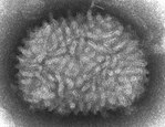 Vaccinia virus Vaccinia virus | ||||||
| Method |  X-RAY DIFFRACTION / SYNCHROTRON / MOLECULAR REPLACEMENT / Resolution: 1.42 Å X-RAY DIFFRACTION / SYNCHROTRON / MOLECULAR REPLACEMENT / Resolution: 1.42 Å | ||||||
 Authors Authors | Matho, M.H. / Zajonc, D.M. | ||||||
 Citation Citation | Journal: J.Virol. / Year: 2012 Title: Structural and Biochemical Characterization of the Vaccinia Virus Envelope Protein D8 and Its Recognition by the Antibody LA5. Authors: Matho, M.H. / Maybeno, M. / Benhnia, M.R. / Becker, D. / Meng, X. / Xiang, Y. / Crotty, S. / Peters, B. / Zajonc, D.M. | ||||||
| History |
|
- Structure visualization
| Structure viewer | Molecule: MolmilJmol/JSmol |
|---|
- Downloads & links
-Download
| PDBx/mmCIF format | 4e9o.cif.gz | 119.3 KB | Display | PDBx/mmCIF format |
|---|---|---|---|---|
| PDB format | pdb4e9o.ent.gz | 92.3 KB | Display | PDB format |
| PDBx/mmJSON format | 4e9o.json.gz | Tree view | PDBx/mmJSON format | |
| Others |  Other downloads Other downloads |
-Validation report
| Summary document | 4e9o_validation.pdf.gz | 436.6 KB | Display | wwPDB validaton report |
|---|---|---|---|---|
| Full document | 4e9o_full_validation.pdf.gz | 438.5 KB | Display | |
| Data in XML | 4e9o_validation.xml.gz | 13.3 KB | Display | |
| Data in CIF | 4e9o_validation.cif.gz | 19 KB | Display | |
| Arichive directory | https://data.pdbj.org/pub/pdb/validation_reports/e9/4e9oftp://data.pdbj.org/pub/pdb/validation_reports/e9/4e9o | HTTPS FTP |
-Related structure data
| Related structure data |  4ebqC  4etqSC S: Starting model for refinement C: citing same article ( |
|---|---|
| Similar structure data |


-Links
 PDBj
PDBj- Assembly
| Deposited unit | 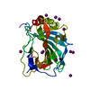
| ||||||||
|---|---|---|---|---|---|---|---|---|---|
| 1 |
| ||||||||
| Unit cell |
|
-Components
| #1: Protein | Mass: 31321.184 Da / Num. of mol.: 1 / Fragment: extracellular domain (UNP residues 1-261) Source method: isolated from a genetically manipulated source Source: (gene. exp.) Vaccinia virus / Strain: Acambis clone 2000 / Gene: VACAC2_124, VACCL3_124, VAC_DPP17_124 / Plasmid: pET22b / Production host:  | ||
|---|---|---|---|
| #2: Chemical | ChemComp-IOD /   Mass: 126.904 Da / Num. of mol.: 12 / Source method: obtained synthetically / Formula: I Mass: 126.904 Da / Num. of mol.: 12 / Source method: obtained synthetically / Formula: I#3: Water | ChemComp-HOH / |  Mass: 18.015 Da / Num. of mol.: 206 / Source method: isolated from a natural source / Formula: H2O Mass: 18.015 Da / Num. of mol.: 206 / Source method: isolated from a natural source / Formula: H2O |
-Experimental details
-Experiment
| Experiment | Method: X-RAY DIFFRACTION / Number of used crystals: 1 |
|---|
- Sample preparation
| Crystal | Density Matthews: 1.8 Å3/Da / Density % sol: 31.71 % |
|---|---|
| Crystal grow | Temperature: 298 K / Method: vapor diffusion Details: 0.2 M sodium iodide, 20% PEG3350, VAPOR DIFFUSION, temperature 298K |
-Data collection
| Diffraction | Mean temperature: 100 K |
|---|---|
| Diffraction source | Source: SYNCHROTRON / Site: SSRL  / Beamline: BL7-1 / Wavelength: 1.100002 Å / Beamline: BL7-1 / Wavelength: 1.100002 Å |
| Detector | Type: ADSC QUANTUM 315r / Detector: CCD / Date: May 23, 2011 / Details: mirrors |
| Radiation | Monochromator: Side scattering I-beam bent single crystal, asymmetric cut 4.9650 degrees Protocol: SINGLE WAVELENGTH / Monochromatic (M) / Laue (L): M / Scattering type: x-ray |
| Radiation wavelength | Wavelength: 1.100002 Å / Relative weight: 1 |
| Reflection | Resolution: 1.42→25.9 Å / Num. all: 40635 / Num. obs: 40635 / % possible obs: 99.3675 % / Observed criterion σ(F): 0 / Observed criterion σ(I): 0 / Redundancy: 2 % / Biso Wilson estimate: 16.2 Å2 / Rsym value: 0.068 / Net I/σ(I): 33.6 |
| Reflection shell | Resolution: 1.42→1.45 Å / Redundancy: 2 % / Mean I/σ(I) obs: 2.7 / Num. unique all: 2880 / Rsym value: 0.63 / % possible all: 100 |
- Processing
| Software |
| ||||||||||||||||||||||||||||||||||||||||||||||||||||||||||||||||||||||||||||||||||||||||||||||||||||
|---|---|---|---|---|---|---|---|---|---|---|---|---|---|---|---|---|---|---|---|---|---|---|---|---|---|---|---|---|---|---|---|---|---|---|---|---|---|---|---|---|---|---|---|---|---|---|---|---|---|---|---|---|---|---|---|---|---|---|---|---|---|---|---|---|---|---|---|---|---|---|---|---|---|---|---|---|---|---|---|---|---|---|---|---|---|---|---|---|---|---|---|---|---|---|---|---|---|---|---|---|---|
| Refinement | Method to determine structure: MOLECULAR REPLACEMENT Starting model: PDB ENTRY 4ETQ Resolution: 1.42→25.9 Å / Cor.coef. Fo:Fc: 0.969 / Cor.coef. Fo:Fc free: 0.962 / SU B: 2.241 / SU ML: 0.044 / Isotropic thermal model: ISOTROPIC / Cross valid method: THROUGHOUT / ESU R: 0.065 / ESU R Free: 0.063 / Stereochemistry target values: MAXIMUM LIKELIHOOD Details: HYDROGENS HAVE BEEN ADDED IN THE RIDING POSITIONS U VALUES : WITH TLS ADDED
| ||||||||||||||||||||||||||||||||||||||||||||||||||||||||||||||||||||||||||||||||||||||||||||||||||||
| Solvent computation | Ion probe radii: 0.8 Å / Shrinkage radii: 0.8 Å / VDW probe radii: 1.4 Å / Solvent model: MASK | ||||||||||||||||||||||||||||||||||||||||||||||||||||||||||||||||||||||||||||||||||||||||||||||||||||
| Displacement parameters | Biso mean: 19.471 Å2
| ||||||||||||||||||||||||||||||||||||||||||||||||||||||||||||||||||||||||||||||||||||||||||||||||||||
| Refinement step | Cycle: LAST / Resolution: 1.42→25.9 Å
| ||||||||||||||||||||||||||||||||||||||||||||||||||||||||||||||||||||||||||||||||||||||||||||||||||||
| Refine LS restraints |
| ||||||||||||||||||||||||||||||||||||||||||||||||||||||||||||||||||||||||||||||||||||||||||||||||||||
| LS refinement shell | Resolution: 1.42→1.457 Å / Total num. of bins used: 20
| ||||||||||||||||||||||||||||||||||||||||||||||||||||||||||||||||||||||||||||||||||||||||||||||||||||
| Refinement TLS params. | Method: refined / Refine-ID: X-RAY DIFFRACTION
| ||||||||||||||||||||||||||||||||||||||||||||||||||||||||||||||||||||||||||||||||||||||||||||||||||||
| Refinement TLS group |
|
