 Movie
Movie Controller

[English] 日本語
 Yorodumi
- PDB-1h6r: The oxidized state of a redox sensitive variant of green fluoresc... -
+ Open data

- Basic information
| Entry | Database: PDB / ID: 1h6r | ||||||
|---|---|---|---|---|---|---|---|
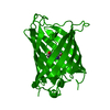
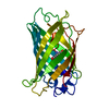
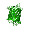
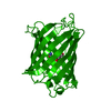
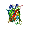
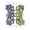
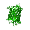
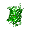
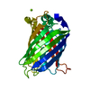
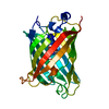
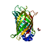
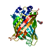
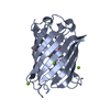
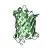
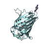
| Title | The oxidized state of a redox sensitive variant of green fluorescent protein | ||||||
 Components Components | GREEN FLUORESCENT PROTEIN | ||||||
 Keywords Keywords | LUMINESCENT PROTEIN / LUMINESCENCE / GREEN FLUORESCENT PROTEIN / YELLOW-EMISSION | ||||||
| Function / homology |  Function and homology information Function and homology information | ||||||
| Biological species |   AEQUOREA VICTORIA (jellyfish) AEQUOREA VICTORIA (jellyfish) | ||||||
| Method | X-RAY DIFFRACTION / SYNCHROTRON / MOLECULAR REPLACEMENT / Resolution: 1.5 Å | ||||||
 Authors Authors | Ostergaard, H. / Henriksen, A. / Hansen, F.G. / Winther, J.R. | ||||||
 Citation Citation | Journal: Embo J. / Year: 2001 Title: Shedding Light on Disulfide Bond Formation: Engineering a Redox Switch in Green Fluorescent Protein Authors: Ostergaard, H. / Henriksen, A. / Hansen, F.G. / Winther, J.R. | ||||||
| History |
| ||||||
| Remark 700 | SHEET DETERMINATION METHOD: DSSP THE SHEETS PRESENTED AS "AA", "BA", "CA" IN CHAIN A, B, C ... SHEET DETERMINATION METHOD: DSSP THE SHEETS PRESENTED AS "AA", "BA", "CA" IN CHAIN A, B, C RESPECTIVELY ON SHEET RECORDS BELOW ARE ACTUALLY AN 11-STRANDED BARREL THIS IS REPRESENTED BY A 12-STRANDED SHEET IN WHICH THE FIRST AND LAST STRANDS ARE IDENTICAL. |
- Structure visualization
| Structure viewer | Molecule: MolmilJmol/JSmol |
|---|
- Downloads & links
-Download
| PDBx/mmCIF format | 1h6r.cif.gz | 168 KB | Display | PDBx/mmCIF format |
|---|---|---|---|---|
| PDB format | pdb1h6r.ent.gz | 133 KB | Display | PDB format |
| PDBx/mmJSON format | 1h6r.json.gz | Tree view | PDBx/mmJSON format | |
| Others |  Other downloads Other downloads |
-Validation report
| Arichive directory | https://data.pdbj.org/pub/pdb/validation_reports/h6/1h6rftp://data.pdbj.org/pub/pdb/validation_reports/h6/1h6r | HTTPS FTP |
|---|
-Related structure data
| Related structure data |  1yfpS S: Starting model for refinement |
|---|---|
| Similar structure data |


-Links
 PDBj
PDBj

- Assembly
| Deposited unit | 
| ||||||||
|---|---|---|---|---|---|---|---|---|---|
| 1 | 
| ||||||||
| 2 | 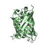
| ||||||||
| 3 | 
| ||||||||
| Unit cell |
|
-Components
| #1: Protein | / GFP Mass: 26944.482 Da / Num. of mol.: 3 / Mutation: YES Source method: isolated from a genetically manipulated source Source: (gene. exp.) AEQUOREA VICTORIA (jellyfish) / Tissue: CIRCUMORAL RING CANAL / Description: SYNTHETIC GENE / Organ: PHOTOGENIC ORGAN / Plasmid: PET24 / Production host:  ESCHERICHIA COLI (E. coli) / Strain (production host): BL21(DE3) / References: UniProt: P42212 ESCHERICHIA COLI (E. coli) / Strain (production host): BL21(DE3) / References: UniProt: P42212#2: Chemical | Chloride  Mass: 35.453 Da / Num. of mol.: 2 / Source method: obtained synthetically / Formula: Cl Mass: 35.453 Da / Num. of mol.: 2 / Source method: obtained synthetically / Formula: Cl#3: Water | ChemComp-HOH / | Water Mass: 18.015 Da / Num. of mol.: 840 / Source method: isolated from a natural source / Formula: H2O Mass: 18.015 Da / Num. of mol.: 840 / Source method: isolated from a natural source / Formula: H2OCompound details | CHAIN A, B, C ENGINEERED MUTATION SER65ALA,CYS48VAL,VAL68LEU, ...CHAIN A, B, C ENGINEERED | Sequence details | MODRES: 1H6R CRO() 66() THE FLUOROPHORE IS GENERATED BY AN AUTOCATALYTIC CYCLIZATION OF THE ...MODRES: 1H6R CRO() 66() THE FLUOROPHOR | |
|---|
-Experimental details
-Experiment
| Experiment | Method: X-RAY DIFFRACTION / Number of used crystals: 1 |
|---|
- Sample preparation
| Crystal | Density Matthews: 2.5 Å3/Da / Density % sol: 49 % | ||||||||||||||||||||||||||||||||||||||||||
|---|---|---|---|---|---|---|---|---|---|---|---|---|---|---|---|---|---|---|---|---|---|---|---|---|---|---|---|---|---|---|---|---|---|---|---|---|---|---|---|---|---|---|---|
| Crystal grow | pH: 8 Details: 100 MM HEPES, PH 8.0, 100 MM MGCL2, AND 14% PEG4000 | ||||||||||||||||||||||||||||||||||||||||||
| Crystal grow | *PLUS Temperature: 22 ℃ / Method: vapor diffusion, hanging drop | ||||||||||||||||||||||||||||||||||||||||||
| Components of the solutions | *PLUS
|
-Data collection
| Diffraction | Mean temperature: 100 K |
|---|---|
| Diffraction source | Source: SYNCHROTRON / Site: MAX II  / Beamline: I711 / Wavelength: 1.0362 / Beamline: I711 / Wavelength: 1.0362 |
| Detector | Type: MARRESEARCH / Detector: IMAGE PLATE / Date: Feb 15, 2001 |
| Radiation | Protocol: SINGLE WAVELENGTH / Monochromatic (M) / Laue (L): M / Scattering type: x-ray |
| Radiation wavelength | Wavelength: 1.0362 Å / Relative weight: 1 |
| Reflection | Resolution: 1.5→78.09 Å / Num. obs: 116321 / % possible obs: 89.1 % / Redundancy: 6.4 % / Biso Wilson estimate: 15.4 Å2 / Rmerge(I) obs: 0.033 / Net I/σ(I): 18.3 |
| Reflection shell | Resolution: 1.5→1.55 Å / Rmerge(I) obs: 0.439 / Mean I/σ(I) obs: 2 / % possible all: 72.9 |
| Reflection | *PLUS Num. measured all: 750960 |
| Reflection shell | *PLUS % possible obs: 72.9 % |
- Processing
| Software |
| ||||||||||||||||||||||||||||||||||||||||||||||||||||||||||||||||||||||||||||||||
|---|---|---|---|---|---|---|---|---|---|---|---|---|---|---|---|---|---|---|---|---|---|---|---|---|---|---|---|---|---|---|---|---|---|---|---|---|---|---|---|---|---|---|---|---|---|---|---|---|---|---|---|---|---|---|---|---|---|---|---|---|---|---|---|---|---|---|---|---|---|---|---|---|---|---|---|---|---|---|---|---|---|
| Refinement | Method to determine structure: MOLECULAR REPLACEMENT Starting model: 1YFP Resolution: 1.5→78.09 Å / Rfactor Rfree error: 0.003 / Data cutoff high absF: 689676.36 / Isotropic thermal model: RESTRAINED / Cross valid method: THROUGHOUT / σ(F): 0 / Stereochemistry target values: MLF Details: A SIGNIFICANT 2FO-FC AND FO-FC DENSITY WAS OBSERVED WITH A CENTER 1.5 ANGSTROM FROM CYS 70 SG AND IT IS POSSIBLE THAT CYS 70 IS IN AN OXIDIZED STATE.
| ||||||||||||||||||||||||||||||||||||||||||||||||||||||||||||||||||||||||||||||||
| Solvent computation | Solvent model: FLAT MODEL / Bsol: 43.493 Å2 / ksol: 0.366229 e/Å3 | ||||||||||||||||||||||||||||||||||||||||||||||||||||||||||||||||||||||||||||||||
| Displacement parameters | Biso mean: 18.9 Å2
| ||||||||||||||||||||||||||||||||||||||||||||||||||||||||||||||||||||||||||||||||
| Refine analyze |
| ||||||||||||||||||||||||||||||||||||||||||||||||||||||||||||||||||||||||||||||||
| Refinement step | Cycle: LAST / Resolution: 1.5→78.09 Å
| ||||||||||||||||||||||||||||||||||||||||||||||||||||||||||||||||||||||||||||||||
| Refine LS restraints |
| ||||||||||||||||||||||||||||||||||||||||||||||||||||||||||||||||||||||||||||||||
| LS refinement shell | Resolution: 1.5→1.59 Å / Rfactor Rfree error: 0.01 / Total num. of bins used: 6
| ||||||||||||||||||||||||||||||||||||||||||||||||||||||||||||||||||||||||||||||||
| Software | *PLUS Name: CNS / Version: 1 / Classification: refinement | ||||||||||||||||||||||||||||||||||||||||||||||||||||||||||||||||||||||||||||||||
| Refine LS restraints | *PLUS
|
