 Movie
Movie Controller

[English] 日本語
 Yorodumi
- PDB-1emb: GREEN FLUORESCENT PROTEIN (GFP) FROM AEQUOREA VICTORIA, GLN 80 RE... -
+ Open data

- Basic information
| Entry | Database: PDB / ID: 1emb | ||||||
|---|---|---|---|---|---|---|---|
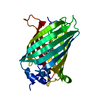
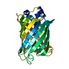
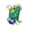
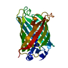
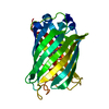
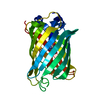
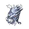
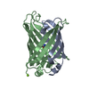
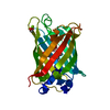
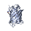
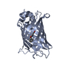
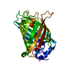
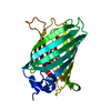
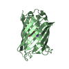
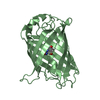
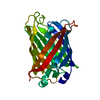
| Title | GREEN FLUORESCENT PROTEIN (GFP) FROM AEQUOREA VICTORIA, GLN 80 REPLACED WITH ARG | ||||||
 Components Components | GREEN FLUORESCENT PROTEIN | ||||||
 Keywords Keywords | FLUORESCENT PROTEIN / BETA-BARREL | ||||||
| Function / homology |  Function and homology information Function and homology informationserine-type endopeptidase inhibitor activity / extracellular space / metal ion binding Similarity search - Function | ||||||
| Biological species |   Aequorea victoria (jellyfish) Aequorea victoria (jellyfish) | ||||||
| Method |  X-RAY DIFFRACTION / MOLECULAR REPLACEMENT / Resolution: 2.13 Å X-RAY DIFFRACTION / MOLECULAR REPLACEMENT / Resolution: 2.13 Å | ||||||
 Authors Authors | Brejc, K. / Sixma, T. | ||||||
 Citation Citation | Journal: Proc.Natl.Acad.Sci.USA / Year: 1997 Title: Structural basis for dual excitation and photoisomerization of the Aequorea victoria green fluorescent protein. Authors: Brejc, K. / Sixma, T.K. / Kitts, P.A. / Kain, S.R. / Tsien, R.Y. / Ormo, M. / Remington, S.J. #1: Journal: Science / Year: 1996Title: Crystal Structure of the Aequorea Victoria Green Fluorescent Protein Authors: Ormo, M. / Cubitt, A.B. / Kallio, K. / Gross, L.A. / Tsien, R.Y. / Remington, S.J. | ||||||
| History |
|
- Structure visualization
| Structure viewer | Molecule: MolmilJmol/JSmol |
|---|
- Downloads & links
-Download
| PDBx/mmCIF format | 1emb.cif.gz | 55.5 KB | Display | PDBx/mmCIF format |
|---|---|---|---|---|
| PDB format | pdb1emb.ent.gz | 43.1 KB | Display | PDB format |
| PDBx/mmJSON format | 1emb.json.gz | Tree view | PDBx/mmJSON format | |
| Others |  Other downloads Other downloads |
-Validation report
| Summary document | 1emb_validation.pdf.gz | 426.2 KB | Display | wwPDB validaton report |
|---|---|---|---|---|
| Full document | 1emb_full_validation.pdf.gz | 434.6 KB | Display | |
| Data in XML | 1emb_validation.xml.gz | 12.4 KB | Display | |
| Data in CIF | 1emb_validation.cif.gz | 16.4 KB | Display | |
| Arichive directory | https://data.pdbj.org/pub/pdb/validation_reports/em/1embftp://data.pdbj.org/pub/pdb/validation_reports/em/1emb | HTTPS FTP |
-Related structure data
| Related structure data |  1emaS S: Starting model for refinement |
|---|---|
| Similar structure data |


-Links
 PDBj
PDBj


- Assembly
| Deposited unit | 
| ||||||||
|---|---|---|---|---|---|---|---|---|---|
| 1 |
| ||||||||
| Unit cell |
|
-Components
| #1: Protein | Mass: 26945.383 Da / Num. of mol.: 1 / Mutation: Q80R Source method: isolated from a genetically manipulated source Details: CHROMOPHORE CONTAINING PROTEIN 65 - 67 DENOTED AS CRO Source: (gene. exp.) Aequorea victoria (jellyfish) / Species (production host): Escherichia coli / Cellular location (production host): CYTOPLASM / Production host:  |
|---|---|
| #2: Water | ChemComp-HOH /  Mass: 18.015 Da / Num. of mol.: 74 / Source method: isolated from a natural source / Formula: H2O Mass: 18.015 Da / Num. of mol.: 74 / Source method: isolated from a natural source / Formula: H2O |
-Experimental details
-Experiment
| Experiment | Method: X-RAY DIFFRACTION / Number of used crystals: 1 |
|---|
- Sample preparation
| Crystal | Density Matthews: 2.09 Å3/Da / Density % sol: 41 % | ||||||||||||||||||||||||||||||||||||||||||||||||
|---|---|---|---|---|---|---|---|---|---|---|---|---|---|---|---|---|---|---|---|---|---|---|---|---|---|---|---|---|---|---|---|---|---|---|---|---|---|---|---|---|---|---|---|---|---|---|---|---|---|
| Crystal grow | pH: 3.8 Details: PROTEIN WAS CRYSTALLIZED FROM 50 MM KH2PO4 AND 20 % (W/V) PEG 8000, PH 3.8. | ||||||||||||||||||||||||||||||||||||||||||||||||
| Crystal grow | *PLUS pH: 8 / Method: vapor diffusion, sitting drop | ||||||||||||||||||||||||||||||||||||||||||||||||
| Components of the solutions | *PLUS
|
-Data collection
| Diffraction | Mean temperature: 295 K |
|---|---|
| Diffraction source | Source: ROTATING ANODE / Type: RIGAKU RUH2R / Wavelength: 1.5418 |
| Detector | Type: MARRESEARCH / Detector: IMAGE PLATE / Date: Sep 1, 1995 |
| Radiation | Monochromator: NI FILTER / Monochromatic (M) / Laue (L): M / Scattering type: x-ray |
| Radiation wavelength | Wavelength: 1.5418 Å / Relative weight: 1 |
| Reflection | Resolution: 2.13→20 Å / Num. obs: 13001 / % possible obs: 91.3 % / Observed criterion σ(I): 3 / Redundancy: 4.8 % / Rsym value: 0.061 / Net I/σ(I): 7.1 |
| Reflection shell | Resolution: 2.13→2.2 Å / Redundancy: 5.3 % / Mean I/σ(I) obs: 3.9 / Rsym value: 0.186 / % possible all: 54.5 |
| Reflection | *PLUS Num. measured all: 60182 / Rmerge(I) obs: 0.061 |
| Reflection shell | *PLUS Lowest resolution: 2.2 Å / % possible obs: 54.5 % / Rmerge(I) obs: 0.186 |
- Processing
| Software |
| ||||||||||||||||||||||||||||||||||||||||||||||||||
|---|---|---|---|---|---|---|---|---|---|---|---|---|---|---|---|---|---|---|---|---|---|---|---|---|---|---|---|---|---|---|---|---|---|---|---|---|---|---|---|---|---|---|---|---|---|---|---|---|---|---|---|
| Refinement | Method to determine structure: MOLECULAR REPLACEMENT Starting model: PDB ENTRY 1EMA Resolution: 2.13→20 Å / Isotropic thermal model: TNT BCORREL / σ(F): 0 / Stereochemistry target values: TNT PROTGEO
| ||||||||||||||||||||||||||||||||||||||||||||||||||
| Solvent computation | Bsol: 145.5 Å2 / ksol: 0.8 e/Å3 | ||||||||||||||||||||||||||||||||||||||||||||||||||
| Refinement step | Cycle: LAST / Resolution: 2.13→20 Å
| ||||||||||||||||||||||||||||||||||||||||||||||||||
| Refine LS restraints |
| ||||||||||||||||||||||||||||||||||||||||||||||||||
| Software | *PLUS Name: TNT / Version: 5E / Classification: refinement | ||||||||||||||||||||||||||||||||||||||||||||||||||
| Refinement | *PLUS Rfactor obs: 0.196 / Rfactor Rfree: 0.254 | ||||||||||||||||||||||||||||||||||||||||||||||||||
| Solvent computation | *PLUS | ||||||||||||||||||||||||||||||||||||||||||||||||||
| Displacement parameters | *PLUS | ||||||||||||||||||||||||||||||||||||||||||||||||||
| Refine LS restraints | *PLUS
|
