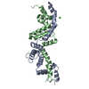
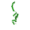
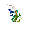
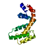
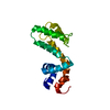
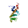
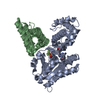
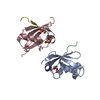
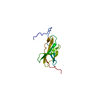
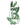
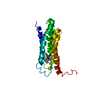
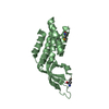
登録情報 データベース : PDB / ID : 2af0タイトル Structure of the Regulator of G-Protein Signaling Domain of RGS2 Regulator of G-protein signaling 2 キーワード / / / / 機能・相同性 分子機能 ドメイン・相同性 構成要素
/ / / / / / / / / / / / / / / / / / / / / / / / / / / / / / / / / / / / / / / / / / / / / / / / / / / / / / / / / / / / / / / 生物種 Homo sapiens (ヒト)手法 / / / 解像度 : 2.3 Å データ登録者 Papagrigoriou, E. / Johannson, C. / Phillips, C. / Smee, C. / Elkins, J.M. / Weigelt, J. / Arrowsmith, C. / Edwards, A. / Sundstrom, M. / Von Delft, F. ...Papagrigoriou, E. / Johannson, C. / Phillips, C. / Smee, C. / Elkins, J.M. / Weigelt, J. / Arrowsmith, C. / Edwards, A. / Sundstrom, M. / Von Delft, F. / Doyle, D.A. / Structural Genomics Consortium (SGC) ジャーナル : Proc.Natl.Acad.Sci.Usa / 年 : 2008タイトル : Structural diversity in the RGS domain and its interaction with heterotrimeric G protein alpha-subunits.著者: Soundararajan, M. / Willard, F.S. / Kimple, A.J. / Turnbull, A.P. / Ball, L.J. / Schoch, G.A. / Gileadi, C. / Fedorov, O.Y. / Dowler, E.F. / Higman, V.A. / Hutsell, S.Q. / Sundstrom, M. / ... 著者 : Soundararajan, M. / Willard, F.S. / Kimple, A.J. / Turnbull, A.P. / Ball, L.J. / Schoch, G.A. / Gileadi, C. / Fedorov, O.Y. / Dowler, E.F. / Higman, V.A. / Hutsell, S.Q. / Sundstrom, M. / Doyle, D.A. / Siderovski, D.P. 履歴 登録 2005年7月25日 登録サイト / 処理サイト 改定 1.0 2005年8月2日 Provider / タイプ 改定 1.1 2008年4月30日 Group 改定 1.2 2011年7月13日 Group 改定 1.3 2024年3月13日 Group / Database referencesカテゴリ chem_comp_atom / chem_comp_bond ... chem_comp_atom / chem_comp_bond / database_2 / struct_ref_seq_dif Item / _database_2.pdbx_database_accession / _struct_ref_seq_dif.details
すべて表示 表示を減らす
 ムービー
ムービー コントローラー


 データを開く
 基本情報
基本情報 要素
要素 キーワード
キーワード 機能・相同性情報
 Homo sapiens (ヒト)
 X線回折 /
  データ登録者
 引用
引用 構造の表示
構造の表示 ダウンロードとリンク
 その他のダウンロード
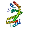


 PDBj
PDBj
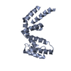
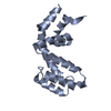
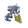
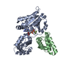
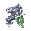
 集合体
集合体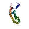

 分子量: 18.015 Da / 分子数: 17 / 由来タイプ: 天然 / 式: H2O
 試料調製
試料調製 / ビームライン: X10SA / 波長: 0.9773 Å
 解析
解析