 Movie
Movie Controller


+ Open data

- Basic information
| Entry | Database: PDB / ID: 4mo7 | ||||||
|---|---|---|---|---|---|---|---|
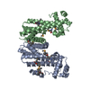
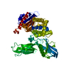
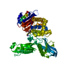
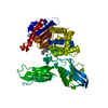
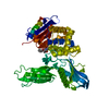
| Title | Crystal structure of superantigen PfiT | ||||||
 Components Components | Transcriptional regulator I2 | ||||||
 Keywords Keywords | TRANSCRIPTION / TetR / Superantigen | ||||||
| Function / homology | Tetracycline Repressor, domain 2 / Tetracycline Repressor; domain 2 / Homeodomain-like / Arc Repressor Mutant, subunit A / Orthogonal Bundle / Mainly Alpha / BETA-MERCAPTOETHANOL / :  Function and homology information Function and homology information | ||||||
| Biological species |  Pseudomonas fluorescens A506 (bacteria) Pseudomonas fluorescens A506 (bacteria) | ||||||
| Method |  X-RAY DIFFRACTION / SYNCHROTRON / SAD / Resolution: 1.701 Å X-RAY DIFFRACTION / SYNCHROTRON / SAD / Resolution: 1.701 Å | ||||||
 Authors Authors | Liu, L.H. / Chen, H. / Li, H.M. | ||||||
 Citation Citation | Journal: Plos Pathog. / Year: 2013 Title: Pfit is a structurally novel Crohn's disease-associated superantigen. Authors: Liu, L. / Chen, H. / Brecher, M.B. / Li, Z. / Wei, B. / Nandi, B. / Zhang, J. / Ling, H. / Winslow, G. / Braun, J. / Li, H. | ||||||
| History |
|
- Structure visualization
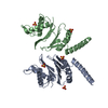
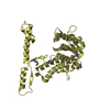
| Structure viewer | Molecule: MolmilJmol/JSmol |
|---|
- Downloads & links
-Download
| PDBx/mmCIF format | 4mo7.cif.gz | 54.9 KB | Display | PDBx/mmCIF format |
|---|---|---|---|---|
| PDB format | pdb4mo7.ent.gz | 39.3 KB | Display | PDB format |
| PDBx/mmJSON format | 4mo7.json.gz | Tree view | PDBx/mmJSON format | |
| Others |  Other downloads Other downloads |
-Validation report
| Arichive directory | https://data.pdbj.org/pub/pdb/validation_reports/mo/4mo7ftp://data.pdbj.org/pub/pdb/validation_reports/mo/4mo7 | HTTPS FTP |
|---|
-Related structure data


-Links
 PDBj
PDBj


- Assembly
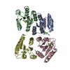
| Deposited unit | 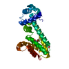
| |||||||||||||||
|---|---|---|---|---|---|---|---|---|---|---|---|---|---|---|---|---|
| 1 | 
| |||||||||||||||
| Unit cell |
| |||||||||||||||
| Components on special symmetry positions |
|
-Components
| #1: Protein | Mass: 23637.109 Da / Num. of mol.: 1 Source method: isolated from a genetically manipulated source Source: (gene. exp.) Pseudomonas fluorescens A506 (bacteria)Gene: pfiT, PflA506_3695 / Plasmid: pGEX-6P-1 / Production host: |
|---|---|
| #2: Chemical | ChemComp-MG /   Mass: 24.305 Da / Num. of mol.: 1 / Source method: obtained synthetically / Formula: Mg Mass: 24.305 Da / Num. of mol.: 1 / Source method: obtained synthetically / Formula: Mg |
| #3: Chemical | ChemComp-BME /   Mass: 78.133 Da / Num. of mol.: 1 / Source method: obtained synthetically / Formula: C2H6OS Mass: 78.133 Da / Num. of mol.: 1 / Source method: obtained synthetically / Formula: C2H6OS |
| #4: Water | ChemComp-HOH /  Mass: 18.015 Da / Num. of mol.: 111 / Source method: isolated from a natural source / Formula: H2O Mass: 18.015 Da / Num. of mol.: 111 / Source method: isolated from a natural source / Formula: H2O |
| Has protein modification | Y |
-Experimental details
-Experiment
| Experiment | Method: X-RAY DIFFRACTION / Number of used crystals: 1 |
|---|
- Sample preparation
| Crystal | Density Matthews: 1.86 Å3/Da / Density % sol: 33.8 % |
|---|---|
| Crystal grow | Temperature: 298 K / Method: vapor diffusion, hanging drop / pH: 8 Details: 4-8% PEG 3350, 0.1 M Tris, 0.2 M ammonium acetate, 5 mM MgCl2, 5 mM DTT, 5% isopropanol, and 5% glycerol, VAPOR DIFFUSION, HANGING DROP, temperature 298K |
-Data collection
| Diffraction | Mean temperature: 100 K |
|---|---|
| Diffraction source | Source: SYNCHROTRON / Site: NSLS  / Beamline: X4A / Wavelength: 1.04023 Å / Beamline: X4A / Wavelength: 1.04023 Å |
| Detector | Type: ADSC QUANTUM 4 / Detector: CCD / Date: Feb 11, 2005 |
| Radiation | Monochromator: mirror / Protocol: SINGLE WAVELENGTH / Monochromatic (M) / Laue (L): M / Scattering type: x-ray |
| Radiation wavelength | Wavelength: 1.04023 Å / Relative weight: 1 |
| Reflection | Resolution: 1.7→50 Å / Num. all: 57293 / Num. obs: 18843 / % possible obs: 99.6 % / Observed criterion σ(F): 1 / Observed criterion σ(I): 1 / Redundancy: 3 % / Rmerge(I) obs: 0.049 / Rsym value: 0.049 / Net I/σ(I): 30.3 |
| Reflection shell | Resolution: 1.7→1.76 Å / Redundancy: 2.8 % / Rmerge(I) obs: 0.556 / Mean I/σ(I) obs: 2.7 / Num. unique all: 1851 / Rsym value: 0.556 / % possible all: 98.8 |
- Processing
| Software |
| ||||||||||||||||||||||||||||||||||||||||||||||||||||||||
|---|---|---|---|---|---|---|---|---|---|---|---|---|---|---|---|---|---|---|---|---|---|---|---|---|---|---|---|---|---|---|---|---|---|---|---|---|---|---|---|---|---|---|---|---|---|---|---|---|---|---|---|---|---|---|---|---|---|
| Refinement | Method to determine structure: SAD / Resolution: 1.701→25.192 Å / SU ML: 0.27 / σ(F): 1.35 / Phase error: 24.75 / Stereochemistry target values: ML
| ||||||||||||||||||||||||||||||||||||||||||||||||||||||||
| Solvent computation | Shrinkage radii: 0.73 Å / VDW probe radii: 1 Å / Solvent model: FLAT BULK SOLVENT MODEL / Bsol: 61.804 Å2 / ksol: 0.404 e/Å3 | ||||||||||||||||||||||||||||||||||||||||||||||||||||||||
| Displacement parameters |
| ||||||||||||||||||||||||||||||||||||||||||||||||||||||||
| Refinement step | Cycle: LAST / Resolution: 1.701→25.192 Å
| ||||||||||||||||||||||||||||||||||||||||||||||||||||||||
| Refine LS restraints |
| ||||||||||||||||||||||||||||||||||||||||||||||||||||||||
| LS refinement shell |
|
