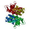
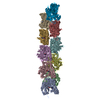
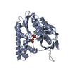
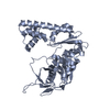
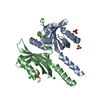
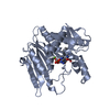
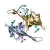
Journal: Science / Year: 2012 Title: A bipolar spindle of antiparallel ParM filaments drives bacterial plasmid segregation. Authors: P Gayathri / T Fujii / J Møller-Jensen / F van den Ent / K Namba / J Löwe / Abstract: To ensure their stable inheritance by daughter cells during cell division, bacterial low-copy-number plasmids make simple DNA segregating machines that use an elongating protein filament between ...To ensure their stable inheritance by daughter cells during cell division, bacterial low-copy-number plasmids make simple DNA segregating machines that use an elongating protein filament between sister plasmids. In the ParMRC system of the Escherichia coli R1 plasmid, ParM, an actinlike protein, forms the spindle between ParRC complexes on sister plasmids. By using a combination of structural work and total internal reflection fluorescence microscopy, we show that ParRC bound and could accelerate growth at only one end of polar ParM filaments, mechanistically resembling eukaryotic formins. The architecture of ParM filaments enabled two ParRC-bound filaments to associate in an antiparallel orientation, forming a bipolar spindle. The spindle elongated as a bundle of at least two antiparallel filaments, thereby pushing two plasmid clusters toward the poles.
In the structure databanks used in Yorodumi, some data are registered as the other names, "COVID-19 virus" and "2019-nCoV". Here are the details of the virus and the list of structure data.
Jan 31, 2019. EMDB accession codes are about to change! (news from PDBe EMDB page)
EMDB accession codes are about to change! (news from PDBe EMDB page)
The allocation of 4 digits for EMDB accession codes will soon come to an end. Whilst these codes will remain in use, new EMDB accession codes will include an additional digit and will expand incrementally as the available range of codes is exhausted. The current 4-digit format prefixed with “EMD-” (i.e. EMD-XXXX) will advance to a 5-digit format (i.e. EMD-XXXXX), and so on. It is currently estimated that the 4-digit codes will be depleted around Spring 2019, at which point the 5-digit format will come into force.
The EM Navigator/Yorodumi systems omit the EMD- prefix.
Related info.:Q: What is EMD? / ID/Accession-code notation in Yorodumi/EM Navigator
Yorodumi is a browser for structure data from EMDB, PDB, SASBDB, etc.
This page is also the successor to EM Navigator detail page, and also detail information page/front-end page for Omokage search.
The word "yorodu" (or yorozu) is an old Japanese word meaning "ten thousand". "mi" (miru) is to see.
Related info.:EMDB / PDB / SASBDB / Comparison of 3 databanks / Yorodumi Search / Aug 31, 2016. New EM Navigator & Yorodumi / Yorodumi Papers / Jmol/JSmol / Function and homology information / Changes in new EM Navigator and Yorodumi
 Movie
Movie Controller

 Yorodumi
 Open data
 Basic information
 Components
 Keywords
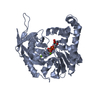
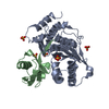
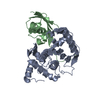
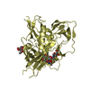
 TRANSPORT PROTEIN / PLASMID SEGREGATION
 Function and homology information
 Authors
 Citation
 Structure visualization
 Downloads & links
 Other downloads
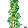


 PDBj
PDBj


 Assembly

 Mass: 506.196 Da / Num. of mol.: 2 / Source method: obtained synthetically / Formula: C10H17N6O12P3 / Comment: AMP-PNP, energy-carrying molecule analogue*YM
 Mass: 24.305 Da / Num. of mol.: 2 / Source method: obtained synthetically / Formula: Mg
 Mass: 18.015 Da / Num. of mol.: 385 / Source method: isolated from a natural source / Formula: H2O
 Sample preparation
 / Beamline: ID14-2 / Wavelength: 0.933
  Processing