 Movie
Movie Controller


+ Open data

- Basic information
| Entry | Database: PDB / ID: 1mwk | ||||||
|---|---|---|---|---|---|---|---|
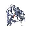
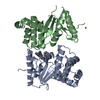
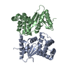
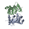
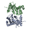
| Title | ParM from plasmid R1 APO form | ||||||
 Components Components | ParM | ||||||
 Keywords Keywords | STRUCTURAL PROTEIN / plasmid / plasmid partition | ||||||
| Function / homology |  Function and homology information Function and homology information | ||||||
| Biological species |  | ||||||
| Method |  X-RAY DIFFRACTION / SYNCHROTRON / MOLECULAR REPLACEMENT / Resolution: 2.3 Å X-RAY DIFFRACTION / SYNCHROTRON / MOLECULAR REPLACEMENT / Resolution: 2.3 Å | ||||||
 Authors Authors | Van den Ent, F. / Moller-Jensen, J. / Amos, L.A. / Gerdes, K. / Lowe, J. | ||||||
 Citation Citation | Journal: EMBO J. / Year: 2002 Title: F-actin-like filaments formed by plasmid segregation protein ParM Authors: Van den Ent, F. / Moller-Jensen, J. / Amos, L.A. / Gerdes, K. / Lowe, J. | ||||||
| History |
|
- Structure visualization
| Structure viewer | Molecule: MolmilJmol/JSmol |
|---|
- Downloads & links
-Download
| PDBx/mmCIF format | 1mwk.cif.gz | 137.2 KB | Display | PDBx/mmCIF format |
|---|---|---|---|---|
| PDB format | pdb1mwk.ent.gz | 108.8 KB | Display | PDB format |
| PDBx/mmJSON format | 1mwk.json.gz | Tree view | PDBx/mmJSON format | |
| Others |  Other downloads Other downloads |
-Validation report
| Arichive directory | https://data.pdbj.org/pub/pdb/validation_reports/mw/1mwkftp://data.pdbj.org/pub/pdb/validation_reports/mw/1mwk | HTTPS FTP |
|---|
-Related structure data


-Links
 PDBj
PDBj- Assembly
| Deposited unit | 
| ||||||||
|---|---|---|---|---|---|---|---|---|---|
| 1 | 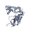
| ||||||||
| 2 | 
| ||||||||
| Unit cell |
|
-Components
| #1: Protein | Mass: 35804.375 Da / Num. of mol.: 2 Source method: isolated from a genetically manipulated source Source: (gene. exp.) #2: Water | ChemComp-HOH / |  Mass: 18.015 Da / Num. of mol.: 282 / Source method: isolated from a natural source / Formula: H2O Mass: 18.015 Da / Num. of mol.: 282 / Source method: isolated from a natural source / Formula: H2O |
|---|
-Experimental details
-Experiment
| Experiment | Method: X-RAY DIFFRACTION / Number of used crystals: 1 |
|---|
- Sample preparation
| Crystal | Density Matthews: 2.61 Å3/Da / Density % sol: 52.8 % | ||||||||||||||||||||||||||||
|---|---|---|---|---|---|---|---|---|---|---|---|---|---|---|---|---|---|---|---|---|---|---|---|---|---|---|---|---|---|
| Crystal grow | Temperature: 291 K / Method: vapor diffusion, sitting drop / pH: 6.5 Details: 1.5M Na3-citrate, 90mM Guanidinium chloride, pH 6.5, VAPOR DIFFUSION, SITTING DROP, temperature 291K | ||||||||||||||||||||||||||||
| Crystal grow | *PLUS Temperature: 19 ℃ | ||||||||||||||||||||||||||||
| Components of the solutions | *PLUS
|
-Data collection
| Diffraction | Mean temperature: 100 K |
|---|---|
| Diffraction source | Source: SYNCHROTRON / Site: ESRF  / Beamline: ID14-4 / Wavelength: 0.9393 Å / Beamline: ID14-4 / Wavelength: 0.9393 Å |
| Detector | Type: ADSC QUANTUM 4 / Detector: CCD / Date: Feb 10, 2002 |
| Radiation | Protocol: SINGLE WAVELENGTH / Monochromatic (M) / Laue (L): M / Scattering type: x-ray |
| Radiation wavelength | Wavelength: 0.9393 Å / Relative weight: 1 |
| Reflection | Resolution: 2.3→40.5 Å / Num. all: 31145 / Num. obs: 31145 / % possible obs: 98.9 % / Observed criterion σ(F): 0 / Observed criterion σ(I): 0 |
| Reflection | *PLUS Redundancy: 3.1 % / Rmerge(I) obs: 0.064 |
| Reflection shell | *PLUS Mean I/σ(I) obs: 3.4 |
- Processing
| Software |
| ||||||||||||||||||||
|---|---|---|---|---|---|---|---|---|---|---|---|---|---|---|---|---|---|---|---|---|---|
| Refinement | Method to determine structure: MOLECULAR REPLACEMENT / Resolution: 2.3→500 Å / σ(F): 0 / Stereochemistry target values: Engh & Huber
| ||||||||||||||||||||
| Refinement step | Cycle: LAST / Resolution: 2.3→500 Å
| ||||||||||||||||||||
| Refinement | *PLUS Lowest resolution: 500 Å | ||||||||||||||||||||
| Solvent computation | *PLUS | ||||||||||||||||||||
| Displacement parameters | *PLUS | ||||||||||||||||||||
| Refine LS restraints | *PLUS
|
