 Movie
Movie Controller


+ Open data

- Basic information
| Entry | Database: EMDB / ID: EMD-1980 | |||||||||
|---|---|---|---|---|---|---|---|---|---|---|
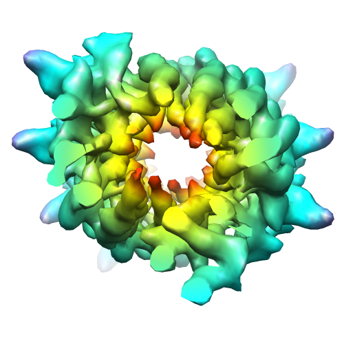
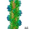
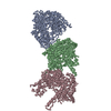
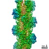
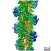
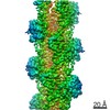
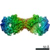
| Title | CryoEM map of ParM filament at 7.2 Angstrom resolution | |||||||||
 Map data Map data | This is an volume file of parM filament | |||||||||
 Sample Sample |
| |||||||||
| Function / homology |  Function and homology information Function and homology information | |||||||||
| Biological species |  | |||||||||
| Method | helical reconstruction / cryo EM / Resolution: 7.2 Å | |||||||||
 Authors Authors | Gayathri P / Fujii T / Moller-Jensen J / van den Ent F / Namba K / Lowe J | |||||||||
 Citation Citation | Journal: Science / Year: 2012 Title: A bipolar spindle of antiparallel ParM filaments drives bacterial plasmid segregation. Authors: P Gayathri / T Fujii / J Møller-Jensen / F van den Ent / K Namba / J Löwe /  Abstract: To ensure their stable inheritance by daughter cells during cell division, bacterial low-copy-number plasmids make simple DNA segregating machines that use an elongating protein filament between ...To ensure their stable inheritance by daughter cells during cell division, bacterial low-copy-number plasmids make simple DNA segregating machines that use an elongating protein filament between sister plasmids. In the ParMRC system of the Escherichia coli R1 plasmid, ParM, an actinlike protein, forms the spindle between ParRC complexes on sister plasmids. By using a combination of structural work and total internal reflection fluorescence microscopy, we show that ParRC bound and could accelerate growth at only one end of polar ParM filaments, mechanistically resembling eukaryotic formins. The architecture of ParM filaments enabled two ParRC-bound filaments to associate in an antiparallel orientation, forming a bipolar spindle. The spindle elongated as a bundle of at least two antiparallel filaments, thereby pushing two plasmid clusters toward the poles. | |||||||||
| History |
|
- Structure visualization
| Movie |
Movie viewer |
|---|---|
| Structure viewer | EM map: SurfViewMolmilJmol/JSmol |
| Supplemental images |
- Downloads & links
-EMDB archive
| Map data | emd_1980.map.gz | 3.6 MB | EMDB map data format | |
|---|---|---|---|---|
| Header (meta data) | emd-1980-v30.xmlemd-1980.xml | 10.6 KB 10.6 KB | Display Display | EMDB header |
| Images |  emd_1980.png emd_1980.png | 170.4 KB | ||
| Archive directory |  http://ftp.pdbj.org/pub/emdb/structures/EMD-1980ftp://ftp.pdbj.org/pub/emdb/structures/EMD-1980 http://ftp.pdbj.org/pub/emdb/structures/EMD-1980ftp://ftp.pdbj.org/pub/emdb/structures/EMD-1980 | HTTPS FTP |
-Related structure data
| Related structure data |  4a6jMC 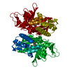 4a61C 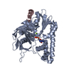 4a62C M: atomic model generated by this map C: citing same article ( |
|---|---|
| Similar structure data |

-Links
| EMDB pages | EMDB (EBI/PDBe) / EMDataResource |
|---|
-Map
| File | Download / File: emd_1980.map.gz / Format: CCP4 / Size: 3.7 MB / Type: IMAGE STORED AS FLOATING POINT NUMBER (4 BYTES) | ||||||||||||||||||||||||||||||||||||||||||||||||||||||||||||||||||||
|---|---|---|---|---|---|---|---|---|---|---|---|---|---|---|---|---|---|---|---|---|---|---|---|---|---|---|---|---|---|---|---|---|---|---|---|---|---|---|---|---|---|---|---|---|---|---|---|---|---|---|---|---|---|---|---|---|---|---|---|---|---|---|---|---|---|---|---|---|---|
| Annotation | This is an volume file of parM filament | ||||||||||||||||||||||||||||||||||||||||||||||||||||||||||||||||||||
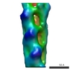
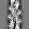
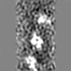
| Projections & slices | Image control
Images are generated by Spider. | ||||||||||||||||||||||||||||||||||||||||||||||||||||||||||||||||||||
| Voxel size | X=Y=Z: 1.64 Å | ||||||||||||||||||||||||||||||||||||||||||||||||||||||||||||||||||||
| Density |
| ||||||||||||||||||||||||||||||||||||||||||||||||||||||||||||||||||||
| Symmetry | Space group: 1 | ||||||||||||||||||||||||||||||||||||||||||||||||||||||||||||||||||||
| Details | EMDB XML:
CCP4 map header:
| ||||||||||||||||||||||||||||||||||||||||||||||||||||||||||||||||||||
 Z (Sec.)
 Y (Row.)
 X (Col.)


-Supplemental data
- Sample components
-Entire : ParM filament
| Entire | Name: ParM filament |
|---|---|
| Components |
|
-Supramolecule #1000: ParM filament
| Supramolecule | Name: ParM filament / type: sample / ID: 1000 / Number unique components: 1 |
|---|
-Macromolecule #1: Plasmid segregation protein parM
| Macromolecule | Name: Plasmid segregation protein parM / type: protein_or_peptide / ID: 1 / Name.synonym: parM / Recombinant expression: Yes |
|---|---|
| Source (natural) | Organism: |
| Molecular weight | Theoretical: 36 KDa |
| Recombinant expression | Organism: |
| Sequence | InterPro: Plasmid segregation protein ParM/StbA |
-Experimental details
-Structure determination
| Method | cryo EM |
|---|---|
 Processing Processing | helical reconstruction |
| Aggregation state | filament |
-Sample preparation
| Buffer | pH: 7.5 Details: 30 mM Tris-HCl, 25 mM KCl, 2 mM MgCl2, 1 mM DTT, pH 7.5 |
|---|---|
| Grid | Details: Quantifoil holey carbon molybdenum grid (R0.6/1.0, Quantifoil Micro Tools GmbH, Jena, Germany) |
| Vitrification | Cryogen name: ETHANE / Chamber humidity: 100 % / Chamber temperature: 50 K / Instrument: OTHER / Details: Vitrification instrument: Vitrobot / Method: Blot for 3.5 seconds before plunging |
- Electron microscopy
| Microscope | JEOL 3200FSC |
|---|---|
| Temperature | Min: 40 K / Max: 60 K / Average: 50 K |
| Specialist optics | Energy filter - Name: Omega filter / Energy filter - Lower energy threshold: 0.0 eV / Energy filter - Upper energy threshold: 10.0 eV |
| Date | Aug 17, 2009 |
| Image recording | Category: CCD / Film or detector model: TVIPS TEMCAM-F415 (4k x 4k) / Digitization - Sampling interval: 15 µm / Number real images: 207 / Average electron dose: 20 e/Å2 / Bits/pixel: 16 |
| Electron beam | Acceleration voltage: 200 kV / Electron source:  FIELD EMISSION GUN FIELD EMISSION GUN |
| Electron optics | Calibrated magnification: 91463 / Illumination mode: FLOOD BEAM / Imaging mode: BRIGHT FIELD / Cs: 1.6 mm / Nominal magnification: 50000 |
| Sample stage | Specimen holder: top entry stage / Specimen holder model: JEOL |
-Image processing
| Final reconstruction | Applied symmetry - Helical parameters - Δz: 23.621 Å Applied symmetry - Helical parameters - Δ&Phi: 164.98 ° Algorithm: OTHER / Resolution.type: BY AUTHOR / Resolution: 7.2 Å / Resolution method: FSC 0.143 CUT-OFF / Software - Name: SPIDER |
|---|---|
| CTF correction | Details: CTFFIND3 Each particle |
