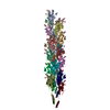
 Movie
Movie Controller


+ Open data

- Basic information
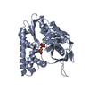
| Entry | Database: PDB / ID: 4a6j | ||||||
|---|---|---|---|---|---|---|---|
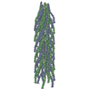
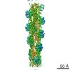
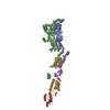
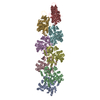
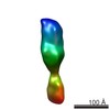
| Title | Structural model of ParM filament based on CryoEM map | ||||||
 Components Components | PLASMID SEGREGATION PROTEIN PARM | ||||||
 Keywords Keywords | TRANSPORT PROTEIN | ||||||
| Function / homology |  Function and homology information Function and homology information | ||||||
| Biological species |  | ||||||
| Method | ELECTRON MICROSCOPY / single particle reconstruction / cryo EM / Resolution: 7.2 Å | ||||||
 Authors Authors | Gayathri, P. / Fujii, T. / Moller-Jensen, J. / Van Den Ent, F. / Namba, K. / Lowe, J. | ||||||
 Citation Citation | Journal: Science / Year: 2012 Title: A bipolar spindle of antiparallel ParM filaments drives bacterial plasmid segregation. Authors: P Gayathri / T Fujii / J Møller-Jensen / F van den Ent / K Namba / J Löwe /  Abstract: To ensure their stable inheritance by daughter cells during cell division, bacterial low-copy-number plasmids make simple DNA segregating machines that use an elongating protein filament between ...To ensure their stable inheritance by daughter cells during cell division, bacterial low-copy-number plasmids make simple DNA segregating machines that use an elongating protein filament between sister plasmids. In the ParMRC system of the Escherichia coli R1 plasmid, ParM, an actinlike protein, forms the spindle between ParRC complexes on sister plasmids. By using a combination of structural work and total internal reflection fluorescence microscopy, we show that ParRC bound and could accelerate growth at only one end of polar ParM filaments, mechanistically resembling eukaryotic formins. The architecture of ParM filaments enabled two ParRC-bound filaments to associate in an antiparallel orientation, forming a bipolar spindle. The spindle elongated as a bundle of at least two antiparallel filaments, thereby pushing two plasmid clusters toward the poles. | ||||||
| History |
|
- Structure visualization
| Movie |
Movie viewer |
|---|---|
| Structure viewer | Molecule: MolmilJmol/JSmol |
- Downloads & links
-Download
| PDBx/mmCIF format | 4a6j.cif.gz | 620 KB | Display | PDBx/mmCIF format |
|---|---|---|---|---|
| PDB format | pdb4a6j.ent.gz | 509.7 KB | Display | PDB format |
| PDBx/mmJSON format | 4a6j.json.gz | Tree view | PDBx/mmJSON format | |
| Others |  Other downloads Other downloads |
-Validation report
| Arichive directory | https://data.pdbj.org/pub/pdb/validation_reports/a6/4a6jftp://data.pdbj.org/pub/pdb/validation_reports/a6/4a6j | HTTPS FTP |
|---|
-Related structure data
| Related structure data | 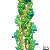 1980MC 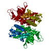 4a61C  4a62C C: citing same article ( M: map data used to model this data |
|---|---|
| Similar structure data |


-Links
 PDBj
PDBj


- Assembly
| Deposited unit | 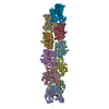
|
|---|---|
| 1 |
|
-Components
| #1: Protein | Mass: 35804.375 Da / Num. of mol.: 10 Source method: isolated from a genetically manipulated source Source: (gene. exp.) #2: Chemical | ChemComp-ANP /   Mass: 506.196 Da / Num. of mol.: 10 / Source method: obtained synthetically / Formula: C10H17N6O12P3 / Comment: AMP-PNP, energy-carrying molecule analogue*YM Mass: 506.196 Da / Num. of mol.: 10 / Source method: obtained synthetically / Formula: C10H17N6O12P3 / Comment: AMP-PNP, energy-carrying molecule analogue*YM#3: Chemical | ChemComp-MG /   Mass: 24.305 Da / Num. of mol.: 10 / Source method: obtained synthetically / Formula: Mg Mass: 24.305 Da / Num. of mol.: 10 / Source method: obtained synthetically / Formula: Mg |
|---|
-Experimental details
-Experiment
| Experiment | Method: ELECTRON MICROSCOPY |
|---|---|
| EM experiment | Aggregation state: FILAMENT / 3D reconstruction method: single particle reconstruction |
- Sample preparation
| Component | Name: PARM FILAMENT / Type: COMPLEX |
|---|---|
| Buffer solution | Name: 30 MM TRIS-HCL, 25 MM KCL, 2 MM MGCL2, 1 MM DTT, 5MM AMPPNP pH: 7.5 Details: 30 MM TRIS-HCL, 25 MM KCL, 2 MM MGCL2, 1 MM DTT, 5MM AMPPNP |
| Specimen | Embedding applied: NO / Shadowing applied: NO / Staining applied: NO / Vitrification applied: YES |
| Specimen support | Details: HOLEY CARBON |
| Vitrification | Instrument: FEI VITROBOT MARK I / Cryogen name: ETHANE / Details: LIQUID ETHENE |
- Electron microscopy imaging
| Microscopy | Model: JEOL 3200FSC / Date: Aug 17, 2009 |
|---|---|
| Electron gun | Electron source:  FIELD EMISSION GUN / Accelerating voltage: 200 kV / Illumination mode: FLOOD BEAM FIELD EMISSION GUN / Accelerating voltage: 200 kV / Illumination mode: FLOOD BEAM |
| Electron lens | Mode: BRIGHT FIELD / Nominal magnification: 50000 X / Calibrated magnification: 91463 X / Cs: 1.6 mm |
| Specimen holder | Temperature: 50 K |
| Image recording | Electron dose: 20 e/Å2 / Film or detector model: TVIPS TEMCAM-F415 (4k x 4k) |
| Image scans | Num. digital images: 207 |
| Radiation wavelength | Relative weight: 1 |
- Processing
| EM software |
| ||||||||||||
|---|---|---|---|---|---|---|---|---|---|---|---|---|---|
| CTF correction | Details: CTFFIND3 EACH PARTICLE | ||||||||||||
| 3D reconstruction | Method: PROJECTION MATCHING / Resolution: 7.2 Å / Actual pixel size: 1.64 Å Details: SUBMISSION BASED ON EXPERIMENTAL DATA FROM EMD-1980. (DEPOSITION ID: 10363). Symmetry type: HELICAL | ||||||||||||
| Atomic model building | Protocol: RIGID BODY FIT / Space: REAL / Details: METHOD--RIGID BODY | ||||||||||||
| Atomic model building | PDB-ID: 4A62 Accession code: 4A62 / Source name: PDB / Type: experimental model | ||||||||||||
| Refinement | Highest resolution: 7.2 Å | ||||||||||||
| Refinement step | Cycle: LAST / Highest resolution: 7.2 Å
|
