 Movie
Movie Controller

[English] 日本語
 Yorodumi
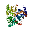
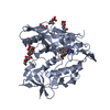
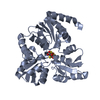
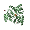
- PDB-5odp: Salinibacter ruber Single-Strand Binding protein D17K D71K mutant -
+ Open data

- Basic information
| Entry | Database: PDB / ID: 5odp | ||||||
|---|---|---|---|---|---|---|---|
| Title | Salinibacter ruber Single-Strand Binding protein D17K D71K mutant | ||||||
 Components Components |
| ||||||
 Keywords Keywords | DNA BINDING PROTEIN / Single-Strand Binding protein | ||||||
| Function / homology |  Function and homology information Function and homology informationnucleoid / single-stranded DNA binding / DNA recombination / DNA replication / DNA repair Similarity search - Function | ||||||
| Biological species |   Salinibacter ruber (bacteria) Salinibacter ruber (bacteria) | ||||||
| Method |  X-RAY DIFFRACTION / SYNCHROTRON / MOLECULAR REPLACEMENT / Resolution: 2.535 Å X-RAY DIFFRACTION / SYNCHROTRON / MOLECULAR REPLACEMENT / Resolution: 2.535 Å | ||||||
 Authors Authors | Pierechod, M. / Rothweiler, U. | ||||||
 Citation Citation | Journal: To Be Published Title: Salinibacter ruber Single-Strand Binding protein D17K D71K mutant Authors: Pierechod, M. / Rothweiler, U. #1: Journal: Patent / Year: 2018Title: Single-strand binding protein Authors: Pierechod, M. / Willassen, N.P. / Rothweiler, U. | ||||||
| History |
|
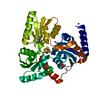
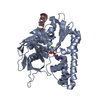
- Structure visualization
| Structure viewer | Molecule: MolmilJmol/JSmol |
|---|
- Downloads & links
-Download
| PDBx/mmCIF format | 5odp.cif.gz | 63.7 KB | Display | PDBx/mmCIF format |
|---|---|---|---|---|
| PDB format | pdb5odp.ent.gz | 43.8 KB | Display | PDB format |
| PDBx/mmJSON format | 5odp.json.gz | Tree view | PDBx/mmJSON format | |
| Others |  Other downloads Other downloads |
-Validation report
| Arichive directory | https://data.pdbj.org/pub/pdb/validation_reports/od/5odpftp://data.pdbj.org/pub/pdb/validation_reports/od/5odp | HTTPS FTP |
|---|
-Related structure data
| Related structure data | 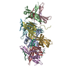 5odnS S: Starting model for refinement |
|---|---|
| Similar structure data |


-Links
 PDBj
PDBj- Assembly
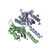
| Deposited unit | 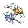
| ||||||||
|---|---|---|---|---|---|---|---|---|---|
| 1 |
| ||||||||
| Unit cell |
|
-Components
| #1: Protein | Mass: 21929.623 Da / Num. of mol.: 2 Source method: isolated from a genetically manipulated source Details: D17K and D71K point mutations Source: (gene. exp.) Salinibacter ruber (strain DSM 13855 / M31) (bacteria)Gene: ssb, SRU_0523 / Production host: #2: DNA chain | Mass: 2996.971 Da / Num. of mol.: 5 / Source method: obtained synthetically / Details: 75 synthetic poly T single stranded DNA / Source: (synth.) Salinibacter ruber (bacteria)#3: Water | ChemComp-HOH / |  Mass: 18.015 Da / Num. of mol.: 4 / Source method: isolated from a natural source / Formula: H2O Mass: 18.015 Da / Num. of mol.: 4 / Source method: isolated from a natural source / Formula: H2O |
|---|
-Experimental details
-Experiment
| Experiment | Method: X-RAY DIFFRACTION / Number of used crystals: 1 |
|---|
- Sample preparation
| Crystal grow | Temperature: 293 K / Method: vapor diffusion, hanging drop / Details: PEG 10k 9.9% |
|---|
-Data collection
| Diffraction | Mean temperature: 100 K |
|---|---|
| Diffraction source | Source: SYNCHROTRON / Site: BESSY  / Beamline: 14.1 / Wavelength: 0.918 Å / Beamline: 14.1 / Wavelength: 0.918 Å |
| Detector | Type: DECTRIS PILATUS 6M / Detector: PIXEL / Date: Jan 24, 2015 |
| Radiation | Protocol: SINGLE WAVELENGTH / Monochromatic (M) / Laue (L): M / Scattering type: x-ray |
| Radiation wavelength | Wavelength: 0.918 Å / Relative weight: 1 |
| Reflection | Resolution: 2.535→50 Å / Num. obs: 11326 / % possible obs: 99.6 % / Redundancy: 12.7 % / CC1/2: 0.999 / Rrim(I) all: 0.122 / Net I/σ(I): 17.59 |
| Reflection shell | Resolution: 2.535→2.69 Å / Mean I/σ(I) obs: 2.19 / Num. unique obs: 1764 / CC1/2: 0.678 / Rrim(I) all: 1.244 / % possible all: 98.9 |
- Processing
| Software |
| |||||||||||||||||||||||||||||||||||
|---|---|---|---|---|---|---|---|---|---|---|---|---|---|---|---|---|---|---|---|---|---|---|---|---|---|---|---|---|---|---|---|---|---|---|---|---|
| Refinement | Method to determine structure: MOLECULAR REPLACEMENT Starting model: 5ODN Resolution: 2.535→46.911 Å / SU ML: 0.33 / Cross valid method: FREE R-VALUE / σ(F): 1.35 / Phase error: 25.47
| |||||||||||||||||||||||||||||||||||
| Solvent computation | Shrinkage radii: 0.9 Å / VDW probe radii: 1.11 Å | |||||||||||||||||||||||||||||||||||
| Refinement step | Cycle: LAST / Resolution: 2.535→46.911 Å
| |||||||||||||||||||||||||||||||||||
| Refine LS restraints |
| |||||||||||||||||||||||||||||||||||
| LS refinement shell |
|
