 Movie
Movie Controller

[English] 日本語
 Yorodumi
- PDB-3hn4: Crystal structure of the NK2 fragment (28-289) of human hepatocyt... -
+ Open data

- Basic information
| Entry | Database: PDB / ID: 3hn4 | ||||||
|---|---|---|---|---|---|---|---|
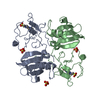
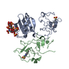
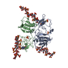
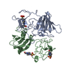
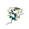
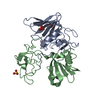
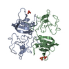
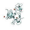
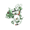
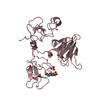
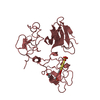
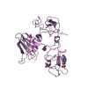
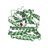
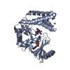
| Title | Crystal structure of the NK2 fragment (28-289) of human hepatocyte growth factor/scatter factor | ||||||
 Components Components | Hepatocyte growth factor | ||||||
 Keywords Keywords | HORMONE / HGF/SF / hormone/growth factor / Disulfide bond / Glycoprotein / Growth factor / Kringle / Serine protease homolog | ||||||
| Function / homology |  Function and homology information Function and homology informationregulation of p38MAPK cascade / regulation of branching involved in salivary gland morphogenesis by mesenchymal-epithelial signaling / Drug-mediated inhibition of MET activation / skeletal muscle cell proliferation / MET activates STAT3 / negative regulation of hydrogen peroxide-mediated programmed cell death / MET interacts with TNS proteins / MET Receptor Activation / hepatocyte growth factor receptor signaling pathway / MET receptor recycling ...regulation of p38MAPK cascade / regulation of branching involved in salivary gland morphogenesis by mesenchymal-epithelial signaling / Drug-mediated inhibition of MET activation / skeletal muscle cell proliferation / MET activates STAT3 / negative regulation of hydrogen peroxide-mediated programmed cell death / MET interacts with TNS proteins / MET Receptor Activation / hepatocyte growth factor receptor signaling pathway / MET receptor recycling / MET activates PTPN11 / MET activates RAP1 and RAC1 / myoblast proliferation / MET activates PI3K/AKT signaling / MET activates PTK2 signaling / positive regulation of DNA biosynthetic process / cellular response to hepatocyte growth factor stimulus / negative regulation of release of cytochrome c from mitochondria / chemoattractant activity / negative regulation of interleukin-6 production / positive regulation of interleukin-10 production / epithelial to mesenchymal transition / positive regulation of osteoblast differentiation / MET activates RAS signaling / negative regulation of peptidyl-serine phosphorylation / negative regulation of extrinsic apoptotic signaling pathway via death domain receptors / Interleukin-7 signaling / cell chemotaxis / negative regulation of autophagy / platelet alpha granule lumen / liver development / epithelial cell proliferation / growth factor activity / cell morphogenesis / Negative regulation of MET activity / negative regulation of inflammatory response / Constitutive Signaling by Aberrant PI3K in Cancer / positive regulation of peptidyl-tyrosine phosphorylation / Platelet degranulation / PIP3 activates AKT signaling / mitotic cell cycle / PI5P, PP2A and IER3 Regulate PI3K/AKT Signaling / RAF/MAP kinase cascade / Interleukin-4 and Interleukin-13 signaling / positive regulation of MAPK cascade / positive regulation of phosphatidylinositol 3-kinase/protein kinase B signal transduction / positive regulation of cell migration / positive regulation of protein phosphorylation / signaling receptor binding / negative regulation of apoptotic process / positive regulation of transcription by RNA polymerase II / extracellular space / extracellular region / membrane / identical protein bindingSimilarity search - Function | ||||||
| Biological species |  Homo sapiens (human) Homo sapiens (human) | ||||||
| Method | X-RAY DIFFRACTION / SYNCHROTRON / MOLECULAR REPLACEMENT / Resolution: 2.6 Å | ||||||
 Authors Authors | Tolbert, W.D. | ||||||
 Citation Citation | Journal: Proc.Natl.Acad.Sci.USA / Year: 2010 Title: Structural basis for agonism and antagonism of hepatocyte growth factor. Authors: Tolbert, W.D. / Daugherty-Holtrop, J. / Gherardi, E. / Vande Woude, G. / Xu, H.E. | ||||||
| History |
|
- Structure visualization
| Structure viewer | Molecule: MolmilJmol/JSmol |
|---|
- Downloads & links
-Download
| PDBx/mmCIF format | 3hn4.cif.gz | 70.6 KB | Display | PDBx/mmCIF format |
|---|---|---|---|---|
| PDB format | pdb3hn4.ent.gz | 50.4 KB | Display | PDB format |
| PDBx/mmJSON format | 3hn4.json.gz | Tree view | PDBx/mmJSON format | |
| Others |  Other downloads Other downloads |
-Validation report
| Arichive directory | https://data.pdbj.org/pub/pdb/validation_reports/hn/3hn4ftp://data.pdbj.org/pub/pdb/validation_reports/hn/3hn4 | HTTPS FTP |
|---|
-Related structure data
| Related structure data |  3hmrC 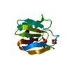 3hmsC 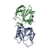 3hmtC 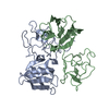 1nk1S S: Starting model for refinement C: citing same article ( |
|---|---|
| Similar structure data |


-Links
 PDBj
PDBj


- Assembly
| Deposited unit | 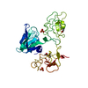
| ||||||||
|---|---|---|---|---|---|---|---|---|---|
| 1 |
| ||||||||
| Unit cell |
|
-Components
| #1: Protein | / Scatter factor / SF / Hepatopoeitin-A / Hepatocyte growth factor alpha chain / Hepatocyte growth ...Scatter factor / SF / Hepatopoeitin-A / Hepatocyte growth factor alpha chain / Hepatocyte growth factor beta chain Mass: 30691.732 Da / Num. of mol.: 1 / Fragment: UNP residues 28-289 / Mutation: K132E, R134E, C214A Source method: isolated from a genetically manipulated source Details: Expressed as a thioredoxin fusion protein and coexpressed with E. coli disulfide bond isomerase C Source: (gene. exp.) Homo sapiens (human) / Gene: HGF, HPTA / Plasmid: pET-Duet1/Trx/DsbC / Production host:  Escherichia coli (E. coli) / Strain (production host): Origami B(DE) / References: UniProt: P14210 Escherichia coli (E. coli) / Strain (production host): Origami B(DE) / References: UniProt: P14210 | ||||
|---|---|---|---|---|---|
| #2: Chemical | ChemComp-EPE / HEPES  Mass: 238.305 Da / Num. of mol.: 1 / Source method: obtained synthetically / Formula: C8H18N2O4S / Comment: pH buffer*YM Mass: 238.305 Da / Num. of mol.: 1 / Source method: obtained synthetically / Formula: C8H18N2O4S / Comment: pH buffer*YM | ||||
| #3: Chemical | 2-Methyl-2,4-pentanediol  Mass: 118.174 Da / Num. of mol.: 2 / Source method: obtained synthetically / Formula: C6H14O2 / Comment: precipitant*YM Mass: 118.174 Da / Num. of mol.: 2 / Source method: obtained synthetically / Formula: C6H14O2 / Comment: precipitant*YM#4: Chemical | Sulfate  Mass: 96.063 Da / Num. of mol.: 2 / Source method: obtained synthetically / Formula: SO4 Mass: 96.063 Da / Num. of mol.: 2 / Source method: obtained synthetically / Formula: SO4#5: Water | ChemComp-HOH / | Water Mass: 18.015 Da / Num. of mol.: 106 / Source method: isolated from a natural source / Formula: H2O Mass: 18.015 Da / Num. of mol.: 106 / Source method: isolated from a natural source / Formula: H2O |
-Experimental details
-Experiment
| Experiment | Method: X-RAY DIFFRACTION / Number of used crystals: 4 |
|---|
- Sample preparation
| Crystal | Density Matthews: 2.52 Å3/Da / Density % sol: 51.13 % |
|---|---|
| Crystal grow | Temperature: 298 K / Method: vapor diffusion, hanging drop / pH: 8 Details: 50 mM Ammonium sulfate, 17-23% PEG 2000 or 4000, 100 mM HEPES pH 8.0, 5% 2-Methyl-2,4-pentanediol, 0.5 mM Beta-octyl glucoside, VAPOR DIFFUSION, HANGING DROP, temperature 298K |
-Data collection
| Diffraction |
| ||||||||||||||||||||||||||||||
|---|---|---|---|---|---|---|---|---|---|---|---|---|---|---|---|---|---|---|---|---|---|---|---|---|---|---|---|---|---|---|---|
| Diffraction source |
| ||||||||||||||||||||||||||||||
| Detector |
| ||||||||||||||||||||||||||||||
| Radiation |
| ||||||||||||||||||||||||||||||
| Radiation wavelength |
| ||||||||||||||||||||||||||||||
| Reflection | Resolution: 2.4→50 Å / Num. all: 13803 / Num. obs: 13803 / % possible obs: 93.7 % / Observed criterion σ(F): 0 / Observed criterion σ(I): 0 / Redundancy: 6.4 % / Rmerge(I) obs: 0.134 / Rsym value: 0.134 / Net I/σ(I): 9.3 | ||||||||||||||||||||||||||||||
| Reflection shell | Resolution: 2.4→2.44 Å / Redundancy: 2.8 % / Rmerge(I) obs: 0.447 / Mean I/σ(I) obs: 1.1 / Rsym value: 0.447 / % possible all: 74.3 |

- Processing
| Software |
| ||||||||||||||||||||||||||||||||||||||||||||||||||||||||||||||||||||||||||||||||||||||||||||||||||||||||||||||||||||||||||||||||||||||||||||||||||||||||||||||||||||||||||
|---|---|---|---|---|---|---|---|---|---|---|---|---|---|---|---|---|---|---|---|---|---|---|---|---|---|---|---|---|---|---|---|---|---|---|---|---|---|---|---|---|---|---|---|---|---|---|---|---|---|---|---|---|---|---|---|---|---|---|---|---|---|---|---|---|---|---|---|---|---|---|---|---|---|---|---|---|---|---|---|---|---|---|---|---|---|---|---|---|---|---|---|---|---|---|---|---|---|---|---|---|---|---|---|---|---|---|---|---|---|---|---|---|---|---|---|---|---|---|---|---|---|---|---|---|---|---|---|---|---|---|---|---|---|---|---|---|---|---|---|---|---|---|---|---|---|---|---|---|---|---|---|---|---|---|---|---|---|---|---|---|---|---|---|---|---|---|---|---|---|---|---|
| Refinement | Method to determine structure: MOLECULAR REPLACEMENT Starting model: PDB entry 1NK1 Resolution: 2.6→41.52 Å / Cor.coef. Fo:Fc: 0.883 / Cor.coef. Fo:Fc free: 0.824 / SU B: 14.594 / SU ML: 0.312 / Isotropic thermal model: RESTRAINED / Cross valid method: THROUGHOUT / ESU R: 2.923 / ESU R Free: 0.446 / Stereochemistry target values: MAXIMUM LIKELIHOOD
| ||||||||||||||||||||||||||||||||||||||||||||||||||||||||||||||||||||||||||||||||||||||||||||||||||||||||||||||||||||||||||||||||||||||||||||||||||||||||||||||||||||||||||
| Solvent computation | Ion probe radii: 0.8 Å / Shrinkage radii: 0.8 Å / VDW probe radii: 1.4 Å / Solvent model: BABINET MODEL WITH MASK | ||||||||||||||||||||||||||||||||||||||||||||||||||||||||||||||||||||||||||||||||||||||||||||||||||||||||||||||||||||||||||||||||||||||||||||||||||||||||||||||||||||||||||
| Displacement parameters | Biso mean: 37.752 Å2
| ||||||||||||||||||||||||||||||||||||||||||||||||||||||||||||||||||||||||||||||||||||||||||||||||||||||||||||||||||||||||||||||||||||||||||||||||||||||||||||||||||||||||||
| Refine analyze |
| ||||||||||||||||||||||||||||||||||||||||||||||||||||||||||||||||||||||||||||||||||||||||||||||||||||||||||||||||||||||||||||||||||||||||||||||||||||||||||||||||||||||||||
| Refinement step | Cycle: LAST / Resolution: 2.6→41.52 Å
| ||||||||||||||||||||||||||||||||||||||||||||||||||||||||||||||||||||||||||||||||||||||||||||||||||||||||||||||||||||||||||||||||||||||||||||||||||||||||||||||||||||||||||
| Refine LS restraints |
| ||||||||||||||||||||||||||||||||||||||||||||||||||||||||||||||||||||||||||||||||||||||||||||||||||||||||||||||||||||||||||||||||||||||||||||||||||||||||||||||||||||||||||
| LS refinement shell | Resolution: 2.6→2.67 Å / Total num. of bins used: 20
| ||||||||||||||||||||||||||||||||||||||||||||||||||||||||||||||||||||||||||||||||||||||||||||||||||||||||||||||||||||||||||||||||||||||||||||||||||||||||||||||||||||||||||
| Xplor file |
|
