 Movie
Movie Controller

[English] 日本語
 Yorodumi
- PDB-2ehb: The structure of the C-terminal domain of the protein kinase AtSO... -
+ Open data

- Basic information
| Entry | Database: PDB / ID: 2ehb | ||||||
|---|---|---|---|---|---|---|---|
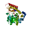
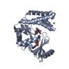
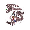
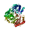
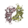
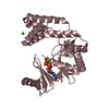
| Title | The structure of the C-terminal domain of the protein kinase AtSOS2 bound to the calcium sensor AtSOS3 | ||||||
 Components Components |
| ||||||
 Keywords Keywords | SIGNALLING PROTEIN/Transferase / protein complex / Ca(II) ions bound to SOS3 (EF-hands 1 and 4) / SOS2 FISL motif / SOS2 phosphatase binding domain (PPI) / SIGNALLING PROTEIN-Transferase COMPLEX | ||||||
| Function / homology |  Function and homology information Function and homology informationhypotonic salinity response / cellular response to salt stress / calcium-dependent protein serine/threonine phosphatase activity / calcineurin complex / plant-type vacuole membrane / intracellular potassium ion homeostasis / detection of calcium ion / response to salt stress / calcium-mediated signaling / kinase binding ...hypotonic salinity response / cellular response to salt stress / calcium-dependent protein serine/threonine phosphatase activity / calcineurin complex / plant-type vacuole membrane / intracellular potassium ion homeostasis / detection of calcium ion / response to salt stress / calcium-mediated signaling / kinase binding / protein kinase activity / non-specific serine/threonine protein kinase / protein serine kinase activity / protein serine/threonine kinase activity / calcium ion binding / signal transduction / ATP binding / nucleus / plasma membrane / cytoplasm Similarity search - Function | ||||||
| Biological species |  | ||||||
| Method |  X-RAY DIFFRACTION / SYNCHROTRON / MOLECULAR REPLACEMENT / Resolution: 2.1 Å X-RAY DIFFRACTION / SYNCHROTRON / MOLECULAR REPLACEMENT / Resolution: 2.1 Å | ||||||
 Authors Authors | Sanchez-Barrena, M.J. | ||||||
 Citation Citation | Journal: Mol.Cell / Year: 2007 Title: The structure of the C-terminal domain of the protein kinase AtSOS2 bound to the calcium sensor AtSOS3 Authors: Sanchez-Barrena, M.J. / Fujii, H. / Angulo, I. / Martinez-Ripoll, M. / Zhu, J.K. / Albert, A. | ||||||
| History |
|
- Structure visualization
| Structure viewer | Molecule: MolmilJmol/JSmol |
|---|
- Downloads & links
-Download
| PDBx/mmCIF format | 2ehb.cif.gz | 81.8 KB | Display | PDBx/mmCIF format |
|---|---|---|---|---|
| PDB format | pdb2ehb.ent.gz | 60.2 KB | Display | PDB format |
| PDBx/mmJSON format | 2ehb.json.gz | Tree view | PDBx/mmJSON format | |
| Others |  Other downloads Other downloads |
-Validation report
| Arichive directory | https://data.pdbj.org/pub/pdb/validation_reports/eh/2ehbftp://data.pdbj.org/pub/pdb/validation_reports/eh/2ehb | HTTPS FTP |
|---|
-Related structure data
| Similar structure data |
|---|


-Links
 PDBj
PDBj- Assembly
| Deposited unit | 
| ||||||||
|---|---|---|---|---|---|---|---|---|---|
| 1 |
| ||||||||
| Unit cell |
|
-Components
| #1: Protein | Mass: 24045.750 Da / Num. of mol.: 1 / Fragment: residues in database 1-207 Source method: isolated from a genetically manipulated source Source: (gene. exp.)  | ||
|---|---|---|---|
| #2: Protein | Mass: 16368.982 Da / Num. of mol.: 1 / Fragment: C-terminal domain (residues 304-446) Source method: isolated from a genetically manipulated source Source: (gene. exp.) References: UniProt: Q9LDI3, non-specific serine/threonine protein kinase | ||
| #3: Chemical |   Mass: 40.078 Da / Num. of mol.: 2 / Source method: obtained synthetically / Formula: Ca Mass: 40.078 Da / Num. of mol.: 2 / Source method: obtained synthetically / Formula: Ca#4: Water | ChemComp-HOH / |  Mass: 18.015 Da / Num. of mol.: 210 / Source method: isolated from a natural source / Formula: H2O Mass: 18.015 Da / Num. of mol.: 210 / Source method: isolated from a natural source / Formula: H2O |
-Experimental details
-Experiment
| Experiment | Method: X-RAY DIFFRACTION / Number of used crystals: 1 |
|---|
- Sample preparation
| Crystal | Density Matthews: 2.22 Å3/Da / Density % sol: 44.72 % |
|---|---|
| Crystal grow | Temperature: 293 K / Method: vapor diffusion, hanging drop / pH: 7.5 Details: drops: 10mg/ml SOS3:C-SOS2, reservoir solution: 20%(w/v) PEG4000, 0.1M Tris-HCl pH 7.5, 0.2M Lithium Sulfate, 3%(v/v) Ethylene Glycol; 1M Guanidine HCl in a ratio of 1:1:0.5, VAPOR ...Details: drops: 10mg/ml SOS3:C-SOS2, reservoir solution: 20%(w/v) PEG4000, 0.1M Tris-HCl pH 7.5, 0.2M Lithium Sulfate, 3%(v/v) Ethylene Glycol; 1M Guanidine HCl in a ratio of 1:1:0.5, VAPOR DIFFUSION, HANGING DROP, temperature 293K |
-Data collection
| Diffraction | Mean temperature: 100 K |
|---|---|
| Diffraction source | Source: SYNCHROTRON / Site: ESRF  / Beamline: ID29 / Wavelength: 0.979 Å / Beamline: ID29 / Wavelength: 0.979 Å |
| Detector | Type: ADSC QUANTUM 315 / Detector: CCD / Date: Feb 16, 2006 Details: The main optical elements are a liquid nitrogen cooled channel-cut silicon monochromator and a cylindrical grazing incidence mirror, the latter which is bent to approximate to a toroidal curvature. |
| Radiation | Monochromator: There is a high resolution Si(311) cut and a lower resolution Si(111) cut Protocol: SINGLE WAVELENGTH / Monochromatic (M) / Laue (L): M / Scattering type: x-ray |
| Radiation wavelength | Wavelength: 0.979 Å / Relative weight: 1 |
| Reflection | Resolution: 2→70.9 Å / Num. all: 25141 / Num. obs: 25141 / % possible obs: 99.3 % / Observed criterion σ(F): 2 / Observed criterion σ(I): 2 / Redundancy: 4.4 % / Rmerge(I) obs: 0.06 / Rsym value: 0.06 / Net I/σ(I): 9.7 |
| Reflection shell | Resolution: 2→2.05 Å / Redundancy: 3.5 % / Rmerge(I) obs: 0.35 / Mean I/σ(I) obs: 2.6 / Rsym value: 0.35 / % possible all: 92.7 |
- Processing
| Software |
| |||||||||||||||||||||||||||||||||||||||||||||||||||||||||||||||||||||||||||||||||||||||||||||||
|---|---|---|---|---|---|---|---|---|---|---|---|---|---|---|---|---|---|---|---|---|---|---|---|---|---|---|---|---|---|---|---|---|---|---|---|---|---|---|---|---|---|---|---|---|---|---|---|---|---|---|---|---|---|---|---|---|---|---|---|---|---|---|---|---|---|---|---|---|---|---|---|---|---|---|---|---|---|---|---|---|---|---|---|---|---|---|---|---|---|---|---|---|---|---|---|---|
| Refinement | Method to determine structure: MOLECULAR REPLACEMENT Starting model: Structure of SOS3 Resolution: 2.1→70.89 Å / Cor.coef. Fo:Fc: 0.935 / Cor.coef. Fo:Fc free: 0.912 / SU B: 6.57 / SU ML: 0.17 / Isotropic thermal model: anisotropic / Cross valid method: THROUGHOUT / σ(F): 2 / σ(I): 2 / ESU R: 0.242 / ESU R Free: 0.201 / Stereochemistry target values: MAXIMUM LIKELIHOOD
| |||||||||||||||||||||||||||||||||||||||||||||||||||||||||||||||||||||||||||||||||||||||||||||||
| Solvent computation | Ion probe radii: 0.8 Å / Shrinkage radii: 0.8 Å / VDW probe radii: 1.2 Å / Solvent model: BABINET MODEL WITH MASK | |||||||||||||||||||||||||||||||||||||||||||||||||||||||||||||||||||||||||||||||||||||||||||||||
| Displacement parameters | Biso mean: 30.969 Å2
| |||||||||||||||||||||||||||||||||||||||||||||||||||||||||||||||||||||||||||||||||||||||||||||||
| Refinement step | Cycle: LAST / Resolution: 2.1→70.89 Å
| |||||||||||||||||||||||||||||||||||||||||||||||||||||||||||||||||||||||||||||||||||||||||||||||
| Refine LS restraints |
| |||||||||||||||||||||||||||||||||||||||||||||||||||||||||||||||||||||||||||||||||||||||||||||||
| LS refinement shell | Resolution: 2.1→2.155 Å / Total num. of bins used: 20
|
