 Movie
Movie Controller

[English] 日本語
 Yorodumi
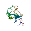
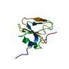
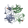
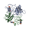
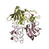
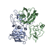
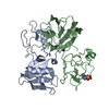
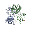
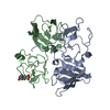
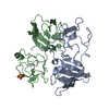
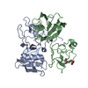
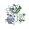
- PDB-1nk1: NK1 FRAGMENT OF HUMAN HEPATOCYTE GROWTH FACTOR/SCATTER FACTOR (HG... -
+ Open data

- Basic information
| Entry | Database: PDB / ID: 1nk1 | ||||||
|---|---|---|---|---|---|---|---|
| Title | NK1 FRAGMENT OF HUMAN HEPATOCYTE GROWTH FACTOR/SCATTER FACTOR (HGF/SF) AT 2.5 ANGSTROM RESOLUTION | ||||||
 Components Components | PROTEIN (HEPATOCYTE GROWTH FACTOR PRECURSOR) | ||||||
 Keywords Keywords | HORMONE/GROWTH FACTOR / HGF/SF / HORMONE-GROWTH FACTOR COMPLEX | ||||||
| Function / homology |  Function and homology information Function and homology informationregulation of p38MAPK cascade / skeletal muscle cell proliferation / regulation of branching involved in salivary gland morphogenesis by mesenchymal-epithelial signaling / Drug-mediated inhibition of MET activation / MET activates STAT3 / negative regulation of hydrogen peroxide-mediated programmed cell death / MET Receptor Activation / MET interacts with TNS proteins / MET receptor recycling / MET activates PTPN11 ...regulation of p38MAPK cascade / skeletal muscle cell proliferation / regulation of branching involved in salivary gland morphogenesis by mesenchymal-epithelial signaling / Drug-mediated inhibition of MET activation / MET activates STAT3 / negative regulation of hydrogen peroxide-mediated programmed cell death / MET Receptor Activation / MET interacts with TNS proteins / MET receptor recycling / MET activates PTPN11 / hepatocyte growth factor receptor signaling pathway / MET activates RAP1 and RAC1 / MET activates PI3K/AKT signaling / MET activates PTK2 signaling / cellular response to hepatocyte growth factor stimulus / positive regulation of DNA biosynthetic process / negative regulation of release of cytochrome c from mitochondria / chemoattractant activity / negative regulation of interleukin-6 production / positive regulation of interleukin-10 production / myoblast proliferation / epithelial to mesenchymal transition / positive regulation of osteoblast differentiation / negative regulation of extrinsic apoptotic signaling pathway via death domain receptors / MET activates RAS signaling / epithelial cell proliferation / platelet alpha granule lumen / Interleukin-7 signaling / negative regulation of autophagy / growth factor activity / cell chemotaxis / liver development / Negative regulation of MET activity / negative regulation of inflammatory response / cell morphogenesis / Constitutive Signaling by Aberrant PI3K in Cancer / Platelet degranulation / PIP3 activates AKT signaling / mitotic cell cycle / PI5P, PP2A and IER3 Regulate PI3K/AKT Signaling / RAF/MAP kinase cascade / Interleukin-4 and Interleukin-13 signaling / positive regulation of MAPK cascade / positive regulation of phosphatidylinositol 3-kinase/protein kinase B signal transduction / positive regulation of cell migration / signaling receptor binding / negative regulation of apoptotic process / positive regulation of transcription by RNA polymerase II / : / extracellular region / membrane / identical protein binding Similarity search - Function | ||||||
| Biological species |  Homo sapiens (human) Homo sapiens (human) | ||||||
| Method |  X-RAY DIFFRACTION / SYNCHROTRON / MOLECULAR REPLACEMENT / Resolution: 2.5 Å X-RAY DIFFRACTION / SYNCHROTRON / MOLECULAR REPLACEMENT / Resolution: 2.5 Å | ||||||
 Authors Authors | Chirgadze, D.Y. / Hepple, J.P. / Zhou, H. / Byrd, R.A. / Blundell, T.L. / Gherardi, E. | ||||||
 Citation Citation | Journal: Nat.Struct.Biol. / Year: 1999 Title: Crystal structure of the NK1 fragment of HGF/SF suggests a novel mode for growth factor dimerization and receptor binding. Authors: Chirgadze, D.Y. / Hepple, J.P. / Zhou, H. / Byrd, R.A. / Blundell, T.L. / Gherardi, E. | ||||||
| History |
|
- Structure visualization
| Structure viewer | Molecule: MolmilJmol/JSmol |
|---|
- Downloads & links
-Download
| PDBx/mmCIF format | 1nk1.cif.gz | 81.1 KB | Display | PDBx/mmCIF format |
|---|---|---|---|---|
| PDB format | pdb1nk1.ent.gz | 61.2 KB | Display | PDB format |
| PDBx/mmJSON format | 1nk1.json.gz | Tree view | PDBx/mmJSON format | |
| Others |  Other downloads Other downloads |
-Validation report
| Arichive directory | https://data.pdbj.org/pub/pdb/validation_reports/nk/1nk1ftp://data.pdbj.org/pub/pdb/validation_reports/nk/1nk1 | HTTPS FTP |
|---|
-Related structure data
-Links
 PDBj
PDBj


- Assembly
| Deposited unit | 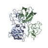
| ||||||||
|---|---|---|---|---|---|---|---|---|---|
| 1 |
| ||||||||
| Unit cell |
| ||||||||
| Noncrystallographic symmetry (NCS) | NCS oper: (Code: given Matrix: (-0.908811, 0.40823, 0.086081), Vector: |
-Components
| #1: Protein | Mass: 21172.273 Da / Num. of mol.: 2 / Fragment: NK1 / Mutation: A29V Source method: isolated from a genetically manipulated source Source: (gene. exp.) Homo sapiens (human) / Description: SYNTHETIC GENE / Cell: FIBROBLAST / Cell line: MRC5 / Cellular location: EXTRACELLULAR / Gene: HEPATOCYTE GROWTH FACTOR/SCATTER FACTOR / Organ: LUNG / Plasmid: PPIC-9K / Cellular location (production host): EXTRACELLULAR / Gene (production host): AOX-1 / Production host:  Pichia pastoris (fungus) / Strain (production host): GS115 / References: UniProt: P14210 Pichia pastoris (fungus) / Strain (production host): GS115 / References: UniProt: P14210#2: Water | ChemComp-HOH / |  Mass: 18.015 Da / Num. of mol.: 59 / Source method: isolated from a natural source / Formula: H2O Mass: 18.015 Da / Num. of mol.: 59 / Source method: isolated from a natural source / Formula: H2OHas protein modification | Y | |
|---|
-Experimental details
-Experiment
| Experiment | Method: X-RAY DIFFRACTION / Number of used crystals: 1 |
|---|
- Sample preparation
| Crystal | Density Matthews: 2.3 Å3/Da / Density % sol: 45 % | ||||||||||||||||||||
|---|---|---|---|---|---|---|---|---|---|---|---|---|---|---|---|---|---|---|---|---|---|
| Crystal grow | pH: 8.7 Details: 18% PEG4000, 0.20M SODIUM ACETATE, 0.15M TRIS, PH 8.5 , pH 8.7 | ||||||||||||||||||||
| Crystal | *PLUS | ||||||||||||||||||||
| Crystal grow | *PLUS Temperature: 25 ℃ / pH: 8.5 / Method: vapor diffusion, hanging drop | ||||||||||||||||||||
| Components of the solutions | *PLUS
|
-Data collection
| Diffraction | Mean temperature: 100 K |
|---|---|
| Diffraction source | Source: SYNCHROTRON / Site: SRS  / Beamline: PX9.5 / Wavelength: 1 / Beamline: PX9.5 / Wavelength: 1 |
| Detector | Type: MARRESEARCH / Detector: IMAGE PLATE / Date: Aug 26, 1997 |
| Radiation | Protocol: SINGLE WAVELENGTH / Monochromatic (M) / Laue (L): M / Scattering type: x-ray |
| Radiation wavelength | Wavelength: 1 Å / Relative weight: 1 |
| Reflection | Resolution: 2.5→30 Å / Num. obs: 13148 / % possible obs: 99.4 % / Redundancy: 3.4 % / Biso Wilson estimate: 41.7 Å2 / Rsym value: 0.042 / Net I/σ(I): 17 |
| Reflection shell | Resolution: 2.5→2.56 Å / Redundancy: 3 % / Rsym value: 0.262 / % possible all: 97.9 |
| Reflection | *PLUS Num. measured all: 44611 / Rmerge(I) obs: 0.042 |
| Reflection shell | *PLUS % possible obs: 97.9 % / Rmerge(I) obs: 0.262 |
- Processing
| Software |
| ||||||||||||||||||||||||||||||||||||||||||||||||||||||||||||||||||||||||||||||||
|---|---|---|---|---|---|---|---|---|---|---|---|---|---|---|---|---|---|---|---|---|---|---|---|---|---|---|---|---|---|---|---|---|---|---|---|---|---|---|---|---|---|---|---|---|---|---|---|---|---|---|---|---|---|---|---|---|---|---|---|---|---|---|---|---|---|---|---|---|---|---|---|---|---|---|---|---|---|---|---|---|---|
| Refinement | Method to determine structure: MOLECULAR REPLACEMENT Starting model: PDB ENTRIES 1PKR AND 2HGF Resolution: 2.5→20 Å / Rfactor Rfree error: 0.013 / Data cutoff high absF: 10000000 / Data cutoff low absF: 0.001 / Isotropic thermal model: RESTRAINED / Cross valid method: THROUGHOUT / σ(F): 0 / Details: BULK SOLVENT MODEL USED
| ||||||||||||||||||||||||||||||||||||||||||||||||||||||||||||||||||||||||||||||||
| Displacement parameters | Biso mean: 41.6 Å2
| ||||||||||||||||||||||||||||||||||||||||||||||||||||||||||||||||||||||||||||||||
| Refine analyze |
| ||||||||||||||||||||||||||||||||||||||||||||||||||||||||||||||||||||||||||||||||
| Refinement step | Cycle: LAST / Resolution: 2.5→20 Å
| ||||||||||||||||||||||||||||||||||||||||||||||||||||||||||||||||||||||||||||||||
| Refine LS restraints |
| ||||||||||||||||||||||||||||||||||||||||||||||||||||||||||||||||||||||||||||||||
| LS refinement shell | Resolution: 2.5→2.66 Å / Rfactor Rfree error: 0.045 / Total num. of bins used: 6
| ||||||||||||||||||||||||||||||||||||||||||||||||||||||||||||||||||||||||||||||||
| Software | *PLUS Name: X-PLOR / Version: 3.851 / Classification: refinement | ||||||||||||||||||||||||||||||||||||||||||||||||||||||||||||||||||||||||||||||||
| Refinement | *PLUS σ(F): 0 / % reflection Rfree: 4.9 % | ||||||||||||||||||||||||||||||||||||||||||||||||||||||||||||||||||||||||||||||||
| Solvent computation | *PLUS | ||||||||||||||||||||||||||||||||||||||||||||||||||||||||||||||||||||||||||||||||
| Displacement parameters | *PLUS Biso mean: 41.6 Å2 | ||||||||||||||||||||||||||||||||||||||||||||||||||||||||||||||||||||||||||||||||
| Refine LS restraints | *PLUS
| ||||||||||||||||||||||||||||||||||||||||||||||||||||||||||||||||||||||||||||||||
| LS refinement shell | *PLUS Rfactor Rfree: 0.474 / % reflection Rfree: 5.1 % / Rfactor Rwork: 0.385 |
