 Movie
Movie Controller


+ Open data

- Basic information
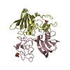
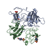
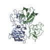
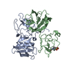
| Entry | Database: PDB / ID: 5csq | ||||||
|---|---|---|---|---|---|---|---|
| Title | The structure of the NK1 fragment of HGF/SF complexed with MOPS | ||||||
 Components Components | Hepatocyte growth factor | ||||||
 Keywords Keywords | HORMONE / HGF/SF / NK1 fragment / fragment based drug discovery / growth factor / cell cycle / new chemical entity | ||||||
| Function / homology |  Function and homology information Function and homology informationregulation of p38MAPK cascade / skeletal muscle cell proliferation / regulation of branching involved in salivary gland morphogenesis by mesenchymal-epithelial signaling / Drug-mediated inhibition of MET activation / MET activates STAT3 / negative regulation of hydrogen peroxide-mediated programmed cell death / MET Receptor Activation / MET interacts with TNS proteins / MET receptor recycling / MET activates PTPN11 ...regulation of p38MAPK cascade / skeletal muscle cell proliferation / regulation of branching involved in salivary gland morphogenesis by mesenchymal-epithelial signaling / Drug-mediated inhibition of MET activation / MET activates STAT3 / negative regulation of hydrogen peroxide-mediated programmed cell death / MET Receptor Activation / MET interacts with TNS proteins / MET receptor recycling / MET activates PTPN11 / hepatocyte growth factor receptor signaling pathway / MET activates RAP1 and RAC1 / MET activates PI3K/AKT signaling / MET activates PTK2 signaling / cellular response to hepatocyte growth factor stimulus / positive regulation of DNA biosynthetic process / negative regulation of release of cytochrome c from mitochondria / chemoattractant activity / negative regulation of interleukin-6 production / myoblast proliferation / positive regulation of interleukin-10 production / epithelial to mesenchymal transition / positive regulation of osteoblast differentiation / negative regulation of extrinsic apoptotic signaling pathway via death domain receptors / MET activates RAS signaling / epithelial cell proliferation / platelet alpha granule lumen / Interleukin-7 signaling / negative regulation of autophagy / growth factor activity / cell chemotaxis / liver development / Negative regulation of MET activity / negative regulation of inflammatory response / cell morphogenesis / Constitutive Signaling by Aberrant PI3K in Cancer / Platelet degranulation / PIP3 activates AKT signaling / mitotic cell cycle / PI5P, PP2A and IER3 Regulate PI3K/AKT Signaling / RAF/MAP kinase cascade / Interleukin-4 and Interleukin-13 signaling / positive regulation of MAPK cascade / positive regulation of phosphatidylinositol 3-kinase/protein kinase B signal transduction / positive regulation of cell migration / signaling receptor binding / negative regulation of apoptotic process / positive regulation of transcription by RNA polymerase II / : / extracellular region / membrane / identical protein binding Similarity search - Function | ||||||
| Biological species |  Homo sapiens (human) Homo sapiens (human) | ||||||
| Method |  X-RAY DIFFRACTION / SYNCHROTRON / MOLECULAR REPLACEMENT / Resolution: 1.95 Å X-RAY DIFFRACTION / SYNCHROTRON / MOLECULAR REPLACEMENT / Resolution: 1.95 Å | ||||||
 Authors Authors | Sigurdardottir, A.G. / Winter, A. / Sobkowicz, A. / Fragai, M. / Chirgadze, D.Y. / Ascher, D.B. / Blundell, T.L. / Gherardi, E. | ||||||
 Citation Citation | Journal: Chem Sci / Year: 2015 Title: Exploring the chemical space of the lysine-binding pocket of the first kringle domain of hepatocyte growth factor/scatter factor (HGF/SF) yields a new class of inhibitors of HGF/SF-MET binding. Authors: Sigurdardottir, A.G. / Winter, A. / Sobkowicz, A. / Fragai, M. / Chirgadze, D. / Ascher, D.B. / Blundell, T.L. / Gherardi, E. | ||||||
| History |
|
- Structure visualization
| Structure viewer | Molecule: MolmilJmol/JSmol |
|---|
- Downloads & links
-Download
| PDBx/mmCIF format | 5csq.cif.gz | 86.8 KB | Display | PDBx/mmCIF format |
|---|---|---|---|---|
| PDB format | pdb5csq.ent.gz | 64.4 KB | Display | PDB format |
| PDBx/mmJSON format | 5csq.json.gz | Tree view | PDBx/mmJSON format | |
| Others |  Other downloads Other downloads |
-Validation report
| Arichive directory | https://data.pdbj.org/pub/pdb/validation_reports/cs/5csqftp://data.pdbj.org/pub/pdb/validation_reports/cs/5csq | HTTPS FTP |
|---|
-Related structure data
| Related structure data | 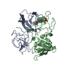 5coeC 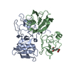 5cp9C 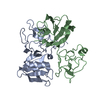 5cs1C  5cs3C 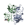 5cs5C 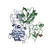 5cs9C 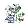 5ct1C 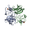 5ct2C 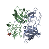 5ct3C  1nk1S S: Starting model for refinement C: citing same article ( |
|---|---|
| Similar structure data |


-Links
 PDBj
PDBj


- Assembly
| Deposited unit | 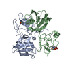
| ||||||||
|---|---|---|---|---|---|---|---|---|---|
| 1 |
| ||||||||
| Unit cell |
|
-Components
| #1: Protein | Mass: 21172.273 Da / Num. of mol.: 2 / Fragment: UNP residues 28-210 / Mutation: A29V Source method: isolated from a genetically manipulated source Source: (gene. exp.) Homo sapiens (human) / Gene: HGF, HPTA / Production host:  Komagataella pastoris CBS 7435 (fungus) / References: UniProt: P14210 Komagataella pastoris CBS 7435 (fungus) / References: UniProt: P14210#2: Chemical |   Mass: 209.263 Da / Num. of mol.: 2 / Source method: obtained synthetically / Formula: C7H15NO4S / Comment: pH buffer*YM Mass: 209.263 Da / Num. of mol.: 2 / Source method: obtained synthetically / Formula: C7H15NO4S / Comment: pH buffer*YM#3: Water | ChemComp-HOH / |  Mass: 18.015 Da / Num. of mol.: 152 / Source method: isolated from a natural source / Formula: H2O Mass: 18.015 Da / Num. of mol.: 152 / Source method: isolated from a natural source / Formula: H2OHas protein modification | Y | |
|---|
-Experimental details
-Experiment
| Experiment | Method: X-RAY DIFFRACTION |
|---|
- Sample preparation
| Crystal | Density Matthews: 2.31 Å3/Da / Density % sol: 46.67 % |
|---|---|
| Crystal grow | Temperature: 293 K / Method: vapor diffusion, hanging drop / Details: 19% PEG 4000, 200 mM NA Acetate, 150 mM Tris / PH range: 7.5 |
-Data collection
| Diffraction | Mean temperature: 100 K |
|---|---|
| Diffraction source | Source: SYNCHROTRON / Site: Diamond  / Beamline: I03 / Wavelength: 0.976 Å / Beamline: I03 / Wavelength: 0.976 Å |
| Detector | Type: DECTRIS PILATUS 6M / Detector: PIXEL / Date: May 27, 2010 |
| Radiation | Protocol: SINGLE WAVELENGTH / Monochromatic (M) / Laue (L): M / Scattering type: x-ray |
| Radiation wavelength | Wavelength: 0.976 Å / Relative weight: 1 |
| Reflection | Resolution: 1.95→50 Å / Num. obs: 27756 / % possible obs: 98.7 % / Redundancy: 3.7 % / Rmerge(I) obs: 0.04 / Rsym value: 0.04 / Net I/σ(I): 20.5 |
| Reflection shell | Resolution: 1.95→2 Å / Redundancy: 3.7 % / Rmerge(I) obs: 0.311 / % possible all: 99.8 |
- Processing
| Software |
| ||||||||||||||||||||||||||||||||||||||||||||||||||||||||||||||||||||||||||||||||||||||||||||||||||||||||||||||||||||||||||||||||||||||||||||||||||||||||||||||||||||||||||||||||||||||
|---|---|---|---|---|---|---|---|---|---|---|---|---|---|---|---|---|---|---|---|---|---|---|---|---|---|---|---|---|---|---|---|---|---|---|---|---|---|---|---|---|---|---|---|---|---|---|---|---|---|---|---|---|---|---|---|---|---|---|---|---|---|---|---|---|---|---|---|---|---|---|---|---|---|---|---|---|---|---|---|---|---|---|---|---|---|---|---|---|---|---|---|---|---|---|---|---|---|---|---|---|---|---|---|---|---|---|---|---|---|---|---|---|---|---|---|---|---|---|---|---|---|---|---|---|---|---|---|---|---|---|---|---|---|---|---|---|---|---|---|---|---|---|---|---|---|---|---|---|---|---|---|---|---|---|---|---|---|---|---|---|---|---|---|---|---|---|---|---|---|---|---|---|---|---|---|---|---|---|---|---|---|---|---|
| Refinement | Method to determine structure: MOLECULAR REPLACEMENT Starting model: 1NK1 Resolution: 1.95→26.29 Å / Cor.coef. Fo:Fc: 0.947 / Cor.coef. Fo:Fc free: 0.923 / SU B: 4.87 / SU ML: 0.139 / Cross valid method: THROUGHOUT / ESU R: 0.201 / ESU R Free: 0.183 / Stereochemistry target values: MAXIMUM LIKELIHOOD / Details: HYDROGENS HAVE BEEN ADDED IN THE RIDING POSITIONS
| ||||||||||||||||||||||||||||||||||||||||||||||||||||||||||||||||||||||||||||||||||||||||||||||||||||||||||||||||||||||||||||||||||||||||||||||||||||||||||||||||||||||||||||||||||||||
| Solvent computation | Ion probe radii: 0.8 Å / Shrinkage radii: 0.8 Å / VDW probe radii: 1.2 Å / Solvent model: MASK | ||||||||||||||||||||||||||||||||||||||||||||||||||||||||||||||||||||||||||||||||||||||||||||||||||||||||||||||||||||||||||||||||||||||||||||||||||||||||||||||||||||||||||||||||||||||
| Displacement parameters | Biso mean: 43.811 Å2
| ||||||||||||||||||||||||||||||||||||||||||||||||||||||||||||||||||||||||||||||||||||||||||||||||||||||||||||||||||||||||||||||||||||||||||||||||||||||||||||||||||||||||||||||||||||||
| Refinement step | Cycle: 1 / Resolution: 1.95→26.29 Å
| ||||||||||||||||||||||||||||||||||||||||||||||||||||||||||||||||||||||||||||||||||||||||||||||||||||||||||||||||||||||||||||||||||||||||||||||||||||||||||||||||||||||||||||||||||||||
| Refine LS restraints |
|