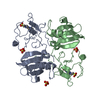
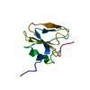
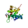
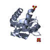
positive regulation of neuron projection regeneration / MET Receptor Activation / Drug-mediated inhibition of MET activation / MET interacts with TNS proteins / MET activates STAT3 / MET activates PI3K/AKT signaling / MET activates PTPN11 / MET activates RAS signaling / MET activates PTK2 signaling / MET activates RAP1 and RAC1 ...positive regulation of neuron projection regeneration / MET Receptor Activation / Drug-mediated inhibition of MET activation / MET interacts with TNS proteins / MET activates STAT3 / MET activates PI3K/AKT signaling / MET activates PTPN11 / MET activates RAS signaling / MET activates PTK2 signaling / MET activates RAP1 and RAC1 / MET receptor recycling / Negative regulation of MET activity / regulation of p38MAPK cascade / PIP3 activates AKT signaling / regulation of branching involved in salivary gland morphogenesis by mesenchymal-epithelial signaling / RAF/MAP kinase cascade / PI5P, PP2A and IER3 Regulate PI3K/AKT Signaling / Platelet degranulation / negative regulation of hydrogen peroxide-mediated programmed cell death / positive regulation of myelination / hepatocyte growth factor receptor signaling pathway / cellular response to hepatocyte growth factor stimulus / positive regulation of DNA biosynthetic process / negative regulation of release of cytochrome c from mitochondria / chemoattractant activity / negative regulation of interleukin-6 production / myoblast proliferation / positive regulation of interleukin-10 production / negative regulation of extrinsic apoptotic signaling pathway via death domain receptors / epithelial cell proliferation / growth factor activity / cell chemotaxis / liver development / negative regulation of inflammatory response / cell morphogenesis / positive regulation of angiogenesis / positive regulation of MAPK cascade / positive regulation of phosphatidylinositol 3-kinase/protein kinase B signal transduction / positive regulation of cell migration / signaling receptor binding / negative regulation of apoptotic process / protein-containing complex binding / proteolysis / : / extracellular region / identical protein binding Similarity search - Function
In the structure databanks used in Yorodumi, some data are registered as the other names, "COVID-19 virus" and "2019-nCoV". Here are the details of the virus and the list of structure data.
Jan 31, 2019. EMDB accession codes are about to change! (news from PDBe EMDB page)
EMDB accession codes are about to change! (news from PDBe EMDB page)
The allocation of 4 digits for EMDB accession codes will soon come to an end. Whilst these codes will remain in use, new EMDB accession codes will include an additional digit and will expand incrementally as the available range of codes is exhausted. The current 4-digit format prefixed with “EMD-” (i.e. EMD-XXXX) will advance to a 5-digit format (i.e. EMD-XXXXX), and so on. It is currently estimated that the 4-digit codes will be depleted around Spring 2019, at which point the 5-digit format will come into force.
The EM Navigator/Yorodumi systems omit the EMD- prefix.
Related info.:Q: What is EMD? / ID/Accession-code notation in Yorodumi/EM Navigator
Yorodumi is a browser for structure data from EMDB, PDB, SASBDB, etc.
This page is also the successor to EM Navigator detail page, and also detail information page/front-end page for Omokage search.
The word "yorodu" (or yorozu) is an old Japanese word meaning "ten thousand". "mi" (miru) is to see.
Related info.:EMDB / PDB / SASBDB / Comparison of 3 databanks / Yorodumi Search / Aug 31, 2016. New EM Navigator & Yorodumi / Yorodumi Papers / Jmol/JSmol / Function and homology information / Changes in new EM Navigator and Yorodumi
 Movie
Movie Controller

 Yorodumi
 Open data
 Basic information
 Components
 Keywords
 Function and homology information
 X-RAY DIFFRACTION /
  Authors
 Citation
 Structure visualization
 Downloads & links
 Other downloads
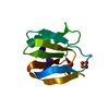


 PDBj
PDBj


 Assembly


 Mass: 96.063 Da / Num. of mol.: 1 / Source method: obtained synthetically / Formula: SO4
 Mass: 18.015 Da / Num. of mol.: 82 / Source method: isolated from a natural source / Formula: H2O
 Sample preparation
 / Beamline: 22-ID / Wavelength: 1 Å
 Processing