 Movie
Movie Controller

[English] 日本語
 Yorodumi
- PDB-1k2d: Crystal structure of the autoimmune MHC class II I-Au complexed w... -
+ Open data

- Basic information
| Entry | Database: PDB / ID: 1k2d | |||||||||
|---|---|---|---|---|---|---|---|---|---|---|
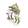
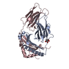
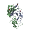
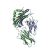
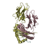
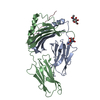
| Title | Crystal structure of the autoimmune MHC class II I-Au complexed with myelin basic protein 1-11 at 2.2A | |||||||||
 Components Components |
| |||||||||
 Keywords Keywords |  IMMUNE SYSTEM / MHC class II / I-Au / H-2u / autoimmune disease / unique register / experimental autoimmune encephalomyelitis / myelin basic protein IMMUNE SYSTEM / MHC class II / I-Au / H-2u / autoimmune disease / unique register / experimental autoimmune encephalomyelitis / myelin basic protein | |||||||||
| Function / homology |  Function and homology information Function and homology informationstructural constituent of myelin sheath / positive regulation of metalloendopeptidase activity / internode region of axon / compact myelin / axon ensheathment / negative regulation of heterotypic cell-cell adhesion / antigen processing and presentation of peptide antigen / EGR2 and SOX10-mediated initiation of Schwann cell myelination / membrane organization / positive regulation of chemokine (C-X-C motif) ligand 2 production ...structural constituent of myelin sheath / positive regulation of metalloendopeptidase activity / internode region of axon / compact myelin / axon ensheathment / negative regulation of heterotypic cell-cell adhesion / antigen processing and presentation of peptide antigen / EGR2 and SOX10-mediated initiation of Schwann cell myelination / membrane organization / positive regulation of chemokine (C-X-C motif) ligand 2 production / positive regulation of T cell differentiation / maintenance of blood-brain barrier / antigen processing and presentation / negative regulation of T cell proliferation / substantia nigra development / myelination / multivesicular body / central nervous system development / cell periphery / sensory perception of sound / response to toxic substance / peptide antigen assembly with MHC class II protein complex / MHC class II protein complex / positive regulation of interleukin-6 production / peptide antigen binding / antigen processing and presentation of exogenous peptide antigen via MHC class II / positive regulation of immune response / MAPK cascade / positive regulation of T cell activation / myelin sheath / MHC class II protein complex binding / late endosome membrane / chemical synaptic transmission / protease binding / adaptive immune response / lysosome / calmodulin binding / early endosome / immune response / lysosomal membrane / external side of plasma membrane / neuronal cell body / synapse / lipid binding / protein-containing complex binding / Golgi apparatus / cell surface / protein-containing complex / nucleus / plasma membrane / cytosolSimilarity search - Function | |||||||||
| Biological species |  Mus musculus (house mouse) Mus musculus (house mouse) | |||||||||
| Method | X-RAY DIFFRACTION / SYNCHROTRON / MOLECULAR REPLACEMENT / Resolution: 2.2 Å | |||||||||
 Authors Authors | He, X.L. / Radu, C. / Ward, E.S. / Garcia, K.C. | |||||||||
 Citation Citation | Journal: IMMUNITY / Year: 2002 Title: Structural snapshot of aberrant antigen presentation linked to autoimmunity: the immunodominant epitope of MBP complexed with I-Au Authors: He, X.L. / Radu, C. / Sidney, J. / Sette, A. / Ward, E.S. / Garcia, K.C. | |||||||||
| History |
| |||||||||
| Remark 999 | SEQUENCE THE C-terminus of the MBP PEPTIDE IS LINKED TO A SYNTHETIC LINKER PEPTIDE, AND ARE Listed ...SEQUENCE THE C-terminus of the MBP PEPTIDE IS LINKED TO A SYNTHETIC LINKER PEPTIDE, AND ARE Listed in the seqres as CHAIN ID P. THE C-terminus of the LINKER PEPTIDE IS LINKED TO THE N-terminus of the BETA CHAIN. THE LINKER PEPTIDE WAS NOT SEEN IN THE ELECTRON DENSITY. |
- Structure visualization
| Structure viewer | Molecule: MolmilJmol/JSmol |
|---|
- Downloads & links
-Download
| PDBx/mmCIF format | 1k2d.cif.gz | 104.9 KB | Display | PDBx/mmCIF format |
|---|---|---|---|---|
| PDB format | pdb1k2d.ent.gz | 78.9 KB | Display | PDB format |
| PDBx/mmJSON format | 1k2d.json.gz | Tree view | PDBx/mmJSON format | |
| Others |  Other downloads Other downloads |
-Validation report
| Arichive directory | https://data.pdbj.org/pub/pdb/validation_reports/k2/1k2dftp://data.pdbj.org/pub/pdb/validation_reports/k2/1k2d | HTTPS FTP |
|---|
-Related structure data
| Similar structure data |
|---|
-Links
 PDBj
PDBj


- Assembly
| Deposited unit | 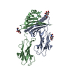
| ||||||||
|---|---|---|---|---|---|---|---|---|---|
| 1 |
| ||||||||
| Unit cell |
|
-Components
| #1: Protein | Mass: 21468.809 Da / Num. of mol.: 1 / Fragment: EXTRACELLULAR ALPHA-1 AND ALPHA-2 DOMAINS Source method: isolated from a genetically manipulated source Source: (gene. exp.) Mus musculus (house mouse) / Production host:  Drosophila melanogaster (fruit fly) / References: UniProt: P14438 Drosophila melanogaster (fruit fly) / References: UniProt: P14438 | ||
|---|---|---|---|
| #2: Protein | Mass: 22495.164 Da / Num. of mol.: 1 / Fragment: EXTRACELLULAR BETA-1 AND BETA-2 DOMAINS Source method: isolated from a genetically manipulated source Source: (gene. exp.) Mus musculus (house mouse) / Production host: Drosophila melanogaster (fruit fly) / References: UniProt: P06344 | ||
| #3: Protein/peptide | Mass: 2319.395 Da / Num. of mol.: 1 / Fragment: 11 residue peptide with 8 residue linker peptide / Source method: obtained synthetically Details: The peptide was chemically synthesized. The sequence of the MBP portion of the peptide is naturally found in Homo sapiens (human). References: GenBank: 14763906, UniProt: P02686*PLUS | ||
| #4: Sugar | N-Acetylglucosamine  Type: D-saccharide, beta linking / Mass: 221.208 Da / Num. of mol.: 3 Type: D-saccharide, beta linking / Mass: 221.208 Da / Num. of mol.: 3Source method: isolated from a genetically manipulated source Formula: C8H15NO6 #5: Water | ChemComp-HOH / | Water Mass: 18.015 Da / Num. of mol.: 450 / Source method: isolated from a natural source / Formula: H2O Mass: 18.015 Da / Num. of mol.: 450 / Source method: isolated from a natural source / Formula: H2O |
-Experimental details
-Experiment
| Experiment | Method: X-RAY DIFFRACTION / Number of used crystals: 1 |
|---|
- Sample preparation
| Crystal | Density Matthews: 2.81 Å3/Da / Density % sol: 56.18 % | ||||||||||||||||||
|---|---|---|---|---|---|---|---|---|---|---|---|---|---|---|---|---|---|---|---|
| Crystal grow | Temperature: 296 K / Method: vapor diffusion, sitting drop / pH: 5.5 Details: MPEG 5000, sodium chloride, citrate, pH 5.5, VAPOR DIFFUSION, SITTING DROP, temperature 296K | ||||||||||||||||||
| Crystal grow | *PLUS pH: 4.5 | ||||||||||||||||||
| Components of the solutions | *PLUS
|
-Data collection
| Diffraction | Mean temperature: 100 K |
|---|---|
| Diffraction source | Source: SYNCHROTRON / Site: SSRL  / Beamline: BL7-1 / Wavelength: 1.08 Å / Beamline: BL7-1 / Wavelength: 1.08 Å |
| Detector | Type: MARRESEARCH / Detector: IMAGE PLATE / Date: Nov 21, 2000 |
| Radiation | Protocol: SINGLE WAVELENGTH / Monochromatic (M) / Laue (L): M / Scattering type: x-ray |
| Radiation wavelength | Wavelength: 1.08 Å / Relative weight: 1 |
| Reflection | Resolution: 2.2→50 Å / Num. all: 26280 / Num. obs: 26280 / % possible obs: 98.3 % / Observed criterion σ(F): 0 / Observed criterion σ(I): 0 / Redundancy: 5.5 % / Rmerge(I) obs: 0.08 / Net I/σ(I): 6.7 |
| Reflection shell | Resolution: 2.2→2.32 Å / Redundancy: 5.6 % / Rmerge(I) obs: 0.448 / Mean I/σ(I) obs: 1.6 / Num. unique all: 3825 / % possible all: 98.7 |
| Reflection | *PLUS Lowest resolution: 50 Å / Num. measured all: 144694 / Rmerge(I) obs: 0.08 |
| Reflection shell | *PLUS Highest resolution: 2.2 Å / Lowest resolution: 2.3 Å / % possible obs: 98.5 % / Rmerge(I) obs: 0.45 |
- Processing
| Software |
| |||||||||||||||||||||||||
|---|---|---|---|---|---|---|---|---|---|---|---|---|---|---|---|---|---|---|---|---|---|---|---|---|---|---|
| Refinement | Method to determine structure: MOLECULAR REPLACEMENT Starting model: PDB ENTRY 1IAKJ Resolution: 2.2→50 Å / Isotropic thermal model: Isotropic / Cross valid method: THROUGHOUT / σ(F): 0 / Stereochemistry target values: Engh & Huber
| |||||||||||||||||||||||||
| Displacement parameters | Biso mean: 50.5 Å2
| |||||||||||||||||||||||||
| Refine analyze |
| |||||||||||||||||||||||||
| Refinement step | Cycle: LAST / Resolution: 2.2→50 Å
| |||||||||||||||||||||||||
| Refine LS restraints |
| |||||||||||||||||||||||||
| LS refinement shell | Resolution: 2.2→2.3 Å / Rfactor Rfree error: 0.029
| |||||||||||||||||||||||||
| Refinement | *PLUS Highest resolution: 2.3 Å / % reflection Rfree: 5 % | |||||||||||||||||||||||||
| Solvent computation | *PLUS | |||||||||||||||||||||||||
| Displacement parameters | *PLUS | |||||||||||||||||||||||||
| Refine LS restraints | *PLUS
|
