 Movie
Movie Controller

[English] 日本語
 Yorodumi
- PDB-2fse: Crystallographic structure of a rheumatoid arthritis MHC suscepti... -
+ Open data

- Basic information
| Entry | Database: PDB / ID: 2fse | ||||||
|---|---|---|---|---|---|---|---|
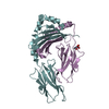
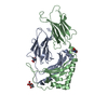
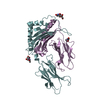
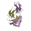
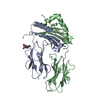
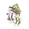
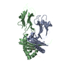
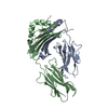
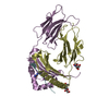
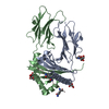
| Title | Crystallographic structure of a rheumatoid arthritis MHC susceptibility allele, HLA-DR1 (DRB1*0101), complexed with the immunodominant determinant of human type II collagen | ||||||
 Components Components |
| ||||||
 Keywords Keywords | IMMUNE SYSTEM / STRUCTURAL PROTEIN / rheumatoid arthritis / HLA-DR1 / collagen type II / antigen presentation | ||||||
| Function / homology |  Function and homology information Function and homology informationcollagen type II trimer / collagen type XI trimer / anterior head development / embryonic skeletal joint morphogenesis / otic vesicle development / Collagen chain trimerization / platelet-derived growth factor binding / extracellular matrix structural constituent conferring tensile strength / regulation of interleukin-4 production / limb bud formation ...collagen type II trimer / collagen type XI trimer / anterior head development / embryonic skeletal joint morphogenesis / otic vesicle development / Collagen chain trimerization / platelet-derived growth factor binding / extracellular matrix structural constituent conferring tensile strength / regulation of interleukin-4 production / limb bud formation / cartilage development involved in endochondral bone morphogenesis / notochord development / regulation of interleukin-10 production / proteoglycan metabolic process / autolysosome membrane / myeloid dendritic cell antigen processing and presentation / antigen processing and presentation of endogenous peptide antigen via MHC class II / Collagen biosynthesis and modifying enzymes / Fibronectin matrix formation / regulation of T-helper cell differentiation / positive regulation of CD4-positive, CD25-positive, alpha-beta regulatory T cell differentiation / tissue homeostasis / MHC class II protein binding / endochondral ossification / positive regulation of CD4-positive, alpha-beta T cell activation / cellular response to BMP stimulus / Signaling by PDGF / antigen processing and presentation of peptide or polysaccharide antigen via MHC class II / positive regulation of T cell mediated immune response to tumor cell / positive regulation of memory T cell differentiation / NCAM1 interactions / inflammatory response to antigenic stimulus / positive regulation of monocyte differentiation / CD4 receptor binding / collagen fibril organization / proteoglycan binding / cartilage development / inner ear morphogenesis / Assembly of collagen fibrils and other multimeric structures / MET activates PTK2 signaling / T-helper 1 type immune response / intermediate filament / cartilage condensation / extracellular matrix structural constituent / roof of mouth development / transport vesicle membrane / Translocation of ZAP-70 to Immunological synapse / Phosphorylation of CD3 and TCR zeta chains / polysaccharide binding / negative regulation of type II interferon production / Collagen degradation / epidermis development / humoral immune response / macrophage differentiation / Generation of second messenger molecules / Non-integrin membrane-ECM interactions / basement membrane / chondrocyte differentiation / Co-inhibition by PD-1 / immunological synapse / ECM proteoglycans / negative regulation of extrinsic apoptotic signaling pathway in absence of ligand / Integrin cell surface interactions / detection of bacterium / negative regulation of T cell proliferation / heart morphogenesis / T cell receptor binding / extrinsic apoptotic signaling pathway in absence of ligand / visual perception / MHC class II antigen presentation / positive regulation of insulin secretion involved in cellular response to glucose stimulus / trans-Golgi network membrane / Developmental Lineage of Pancreatic Ductal Cells / skeletal system development / central nervous system development / lumenal side of endoplasmic reticulum membrane / protein tetramerization / negative regulation of inflammatory response to antigenic stimulus / sensory perception of sound / ER to Golgi transport vesicle membrane / clathrin-coated endocytic vesicle membrane / peptide antigen assembly with MHC class II protein complex / MHC class II protein complex / positive regulation of T cell mediated cytotoxicity / structural constituent of cytoskeleton / antigen processing and presentation of exogenous peptide antigen via MHC class II / positive regulation of immune response / peptide antigen binding / cognition / positive regulation of T cell activation / Interferon gamma signaling / Immunoregulatory interactions between a Lymphoid and a non-Lymphoid cell / endocytic vesicle membrane / MHC class II protein complex binding / T cell receptor signaling pathway / late endosome membrane / Downstream TCR signaling / extracellular matrix / regulation of gene expression / early endosome membrane Similarity search - Function | ||||||
| Biological species |  Homo sapiens (human) Homo sapiens (human) | ||||||
| Method |  X-RAY DIFFRACTION / SYNCHROTRON / MOLECULAR REPLACEMENT / Resolution: 3.1 Å X-RAY DIFFRACTION / SYNCHROTRON / MOLECULAR REPLACEMENT / Resolution: 3.1 Å | ||||||
 Authors Authors | Ivey, R.A. / Rosloniec, E.F. / Whittington, K.B. / Kang, A.H. / Park, H.W. | ||||||
 Citation Citation | Journal: J.Immunol. / Year: 2006 Title: Crystallographic Structure of a Rheumatoid Arthritis MHC Susceptibility Allele, HLA-DR1 (DRB1*0101), Complexed with the Immunodominant Determinant of Human Type II Collagen. Authors: Rosloniec, E.F. / Ivey, R.A. / Whittington, K.B. / Kang, A.H. / Park, H.W. | ||||||
| History |
| ||||||
| Remark 999 | SEQUENCE THERE WAS NO SEQUENCE DATABASE REFERENCE AT THE TIME OF PROCESSING FOR THE MHC CHAINS. |
- Structure visualization
| Structure viewer | Molecule: MolmilJmol/JSmol |
|---|
- Downloads & links
-Download
| PDBx/mmCIF format | 2fse.cif.gz | 160.7 KB | Display | PDBx/mmCIF format |
|---|---|---|---|---|
| PDB format | pdb2fse.ent.gz | 128.7 KB | Display | PDB format |
| PDBx/mmJSON format | 2fse.json.gz | Tree view | PDBx/mmJSON format | |
| Others |  Other downloads Other downloads |
-Validation report
| Arichive directory | https://data.pdbj.org/pub/pdb/validation_reports/fs/2fseftp://data.pdbj.org/pub/pdb/validation_reports/fs/2fse | HTTPS FTP |
|---|
-Related structure data
| Similar structure data |
|---|
-Links
 PDBj
PDBj


- Assembly
| Deposited unit | 
| ||||||||
|---|---|---|---|---|---|---|---|---|---|
| 1 | 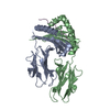
| ||||||||
| 2 | 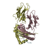
| ||||||||
| Unit cell |
|
-Components
| #1: Protein | Mass: 20711.234 Da / Num. of mol.: 2 Source method: isolated from a genetically manipulated source Details: chains A and C form a chimera with chains B and D, respectively Source: (gene. exp.) Homo sapiens (human) / Plasmid: pRmHA-3 / Production host:  #2: Protein | Mass: 21909.436 Da / Num. of mol.: 2 Source method: isolated from a genetically manipulated source Details: chains B and D form a chimera with chains A and C, respectively Source: (gene. exp.) #3: Protein/peptide | Mass: 1360.471 Da / Num. of mol.: 2 Source method: isolated from a genetically manipulated source Source: (gene. exp.) Homo sapiens (human) / Gene: COL2A1 / Plasmid: pRmHA-3 / Production host: #4: Water | ChemComp-HOH / |  Mass: 18.015 Da / Num. of mol.: 31 / Source method: isolated from a natural source / Formula: H2O Mass: 18.015 Da / Num. of mol.: 31 / Source method: isolated from a natural source / Formula: H2OHas protein modification | Y | |
|---|
-Experimental details
-Experiment
| Experiment | Method: X-RAY DIFFRACTION |
|---|
- Sample preparation
| Crystal | Density Matthews: 3.21 Å3/Da / Density % sol: 61.7 % |
|---|---|
| Crystal grow | Temperature: 293 K / Method: vapor diffusion, hanging drop Details: 20% polyethylene glycol 3,350 and 200 mM ammonium chloride, VAPOR DIFFUSION, HANGING DROP, temperature 293K |
-Data collection
| Diffraction | Mean temperature: 77 K |
|---|---|
| Diffraction source | Source: SYNCHROTRON / Site: APS  / Beamline: 22-ID / Wavelength: 1 Å / Beamline: 22-ID / Wavelength: 1 Å |
| Detector | Detector: CCD |
| Radiation | Protocol: SINGLE WAVELENGTH / Monochromatic (M) / Laue (L): M / Scattering type: x-ray |
| Radiation wavelength | Wavelength: 1 Å / Relative weight: 1 |
| Reflection | Resolution: 3.1→91.287 Å / Num. all: 21330 / Num. obs: 20446 / % possible obs: 82.4 % / Observed criterion σ(F): 2 / Observed criterion σ(I): 2 / Redundancy: 2.9 % / Limit h max: 19 / Limit h min: 0 / Limit k max: 35 / Limit k min: 0 / Limit l max: 55 / Limit l min: 0 / Rsym value: 0.099 / Net I/σ(I): 11.4 |
| Reflection shell | Resolution: 3.1→3.21 Å / Redundancy: 2.9 % / Mean I/σ(I) obs: 2.9 / Num. unique all: 2046 / Rsym value: 0.382 / % possible all: 96.7 |
- Processing
| Software |
| ||||||||||||||||
|---|---|---|---|---|---|---|---|---|---|---|---|---|---|---|---|---|---|
| Refinement | Method to determine structure: MOLECULAR REPLACEMENT / Resolution: 3.1→20 Å / σ(F): 1936
| ||||||||||||||||
| Solvent computation | Bsol: 15.01 Å2 | ||||||||||||||||
| Displacement parameters | Biso mean: 26.877 Å2
| ||||||||||||||||
| Refinement step | Cycle: LAST / Resolution: 3.1→20 Å
| ||||||||||||||||
| Xplor file |
|
