 Movie
Movie Controller

[English] 日本語
 Yorodumi
- PDB-1bx2: CRYSTAL STRUCTURE OF HLA-DR2 (DRA*0101,DRB1*1501) COMPLEXED WITH ... -
+ Open data

- Basic information
| Entry | Database: PDB / ID: 1bx2 | ||||||
|---|---|---|---|---|---|---|---|
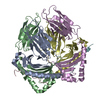
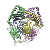
| Title | CRYSTAL STRUCTURE OF HLA-DR2 (DRA*0101,DRB1*1501) COMPLEXED WITH A PEPTIDE FROM HUMAN MYELIN BASIC PROTEIN | ||||||
 Components Components | (PROTEIN (HLA-DR2)) x 3 | ||||||
 Keywords Keywords | IMMUNE SYSTEM / HLA-DR2 / MYELIN BASIC PROTEIN / MULTIPLE SCLEROSIS / AUTOIMMUNITY | ||||||
| Function / homology |  Function and homology information Function and homology informationstructural constituent of myelin sheath / compact myelin / internode region of axon / myelin assembly / axon ensheathment / regulation of interleukin-4 production / regulation of interleukin-10 production / autolysosome membrane / myeloid dendritic cell antigen processing and presentation / antigen processing and presentation of endogenous peptide antigen via MHC class II ...structural constituent of myelin sheath / compact myelin / internode region of axon / myelin assembly / axon ensheathment / regulation of interleukin-4 production / regulation of interleukin-10 production / autolysosome membrane / myeloid dendritic cell antigen processing and presentation / antigen processing and presentation of endogenous peptide antigen via MHC class II / regulation of T-helper cell differentiation / positive regulation of CD4-positive, CD25-positive, alpha-beta regulatory T cell differentiation / positive regulation of CD4-positive, alpha-beta T cell activation / EGR2 and SOX10-mediated initiation of Schwann cell myelination / antigen processing and presentation of peptide or polysaccharide antigen via MHC class II / positive regulation of T cell mediated immune response to tumor cell / membrane organization / positive regulation of memory T cell differentiation / positive regulation of monocyte differentiation / inflammatory response to antigenic stimulus / CD4 receptor binding / T-helper 1 type immune response / intermediate filament / transport vesicle membrane / Translocation of ZAP-70 to Immunological synapse / Phosphorylation of CD3 and TCR zeta chains / polysaccharide binding / negative regulation of type II interferon production / humoral immune response / Generation of second messenger molecules / macrophage differentiation / Co-inhibition by PD-1 / immunological synapse / epidermis development / detection of bacterium / negative regulation of T cell proliferation / T cell receptor binding / myelination / MHC class II antigen presentation / positive regulation of insulin secretion involved in cellular response to glucose stimulus / trans-Golgi network membrane / cell periphery / central nervous system development / lumenal side of endoplasmic reticulum membrane / protein tetramerization / negative regulation of inflammatory response to antigenic stimulus / ER to Golgi transport vesicle membrane / clathrin-coated endocytic vesicle membrane / sensory perception of sound / peptide antigen assembly with MHC class II protein complex / MHC class II protein complex / positive regulation of T cell mediated cytotoxicity / structural constituent of cytoskeleton / antigen processing and presentation of exogenous peptide antigen via MHC class II / positive regulation of immune response / peptide antigen binding / cognition / response to toxic substance / positive regulation of T cell activation / Interferon gamma signaling / endocytic vesicle membrane / MHC class II protein complex binding / myelin sheath / late endosome membrane / Downstream TCR signaling / T cell receptor signaling pathway / protease binding / early endosome membrane / chemical synaptic transmission / adaptive immune response / calmodulin binding / lysosome / immune response / Golgi membrane / external side of plasma membrane / lysosomal membrane / neuronal cell body / lipid binding / synapse / cell surface / signal transduction / protein-containing complex / : / extracellular exosome / membrane / nucleus / plasma membrane / cytosol Similarity search - Function | ||||||
| Biological species |  Homo sapiens (human) Homo sapiens (human) | ||||||
| Method |  X-RAY DIFFRACTION / SYNCHROTRON / MOLECULAR REPLACEMENT / Resolution: 2.6 Å X-RAY DIFFRACTION / SYNCHROTRON / MOLECULAR REPLACEMENT / Resolution: 2.6 Å | ||||||
 Authors Authors | Smith, K.J. / Pyrdol, J. / Gauthier, L. / Wiley, D.C. / Wucherpfennig, K. | ||||||
 Citation Citation | Journal: J.Exp.Med. / Year: 1998 Title: Crystal structure of HLA-DR2 (DRA*0101, DRB1*1501) complexed with a peptide from human myelin basic protein. Authors: Smith, K.J. / Pyrdol, J. / Gauthier, L. / Wiley, D.C. / Wucherpfennig, K.W. #1: Journal: Cell(Cambridge,Mass.) / Year: 1995Title: Molecular Mimicry in T Cell Mediated Autoimmunity:Viral Peptides Activate Human T Cell Clones Specific for Myelin Basic Protein Authors: Wucherpfennig, K. / Strominger, J.L. | ||||||
| History |
|
- Structure visualization
| Structure viewer | Molecule: MolmilJmol/JSmol |
|---|
- Downloads & links
-Download
| PDBx/mmCIF format | 1bx2.cif.gz | 166 KB | Display | PDBx/mmCIF format |
|---|---|---|---|---|
| PDB format | pdb1bx2.ent.gz | 132.1 KB | Display | PDB format |
| PDBx/mmJSON format | 1bx2.json.gz | Tree view | PDBx/mmJSON format | |
| Others |  Other downloads Other downloads |
-Validation report
| Arichive directory | https://data.pdbj.org/pub/pdb/validation_reports/bx/1bx2ftp://data.pdbj.org/pub/pdb/validation_reports/bx/1bx2 | HTTPS FTP |
|---|
-Related structure data
| Related structure data | 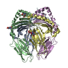 1dlhS S: Starting model for refinement |
|---|---|
| Similar structure data |


-Links
 PDBj
PDBj


- Assembly
| Deposited unit | 
| ||||||||
|---|---|---|---|---|---|---|---|---|---|
| 1 |
| ||||||||
| 2 | 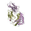
| ||||||||
| 3 | 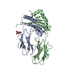
| ||||||||
| Unit cell |
|
-Components
| #1: Protein | Mass: 20971.670 Da / Num. of mol.: 2 / Fragment: EXTRACELLULAR DOMAINS ALPHA 1, ALPHA 2 Source method: isolated from a genetically manipulated source Source: (gene. exp.) Homo sapiens (human) / Cellular location (production host): SECRETED / Production host:  unidentified baculovirus / References: UniProt: P01903 unidentified baculovirus / References: UniProt: P01903#2: Protein | Mass: 22316.930 Da / Num. of mol.: 2 / Fragment: EXTRACELLULAR DOMAINS BETA 1, BETA 2 / Source method: isolated from a natural source / Source: (natural) Homo sapiens (human) / References: UniProt: P04229, UniProt: P01911*PLUS#3: Protein/peptide | Mass: 1800.086 Da / Num. of mol.: 2 / Fragment: PEPTIDE FROM HUMAN MYELIN BASIC PROTEIN / Source method: isolated from a natural source Details: MYELIN BASIC PROTEIN PEPTIDE WAS COVALENTLY LINKED TO THE N-TERMINUS OF THE HLA-DR2 BETA CHAIN Source: (natural) Homo sapiens (human) / References: UniProt: P02686*PLUS#4: Sugar |   Type: D-saccharide, beta linking / Mass: 221.208 Da / Num. of mol.: 2 Type: D-saccharide, beta linking / Mass: 221.208 Da / Num. of mol.: 2Source method: isolated from a genetically manipulated source Formula: C8H15NO6 #5: Water | ChemComp-HOH / |  Mass: 18.015 Da / Num. of mol.: 48 / Source method: isolated from a natural source / Formula: H2O Mass: 18.015 Da / Num. of mol.: 48 / Source method: isolated from a natural source / Formula: H2OHas protein modification | Y | |
|---|
-Experimental details
-Experiment
| Experiment | Method: X-RAY DIFFRACTION / Number of used crystals: 1 |
|---|
- Sample preparation
| Crystal | Density Matthews: 3.2 Å3/Da / Density % sol: 62 % | |||||||||||||||||||||||||
|---|---|---|---|---|---|---|---|---|---|---|---|---|---|---|---|---|---|---|---|---|---|---|---|---|---|---|
| Crystal grow | pH: 3.5 / Details: 15-18% PEG6000 100MM GLYCINE PH3.5 | |||||||||||||||||||||||||
| Crystal grow | *PLUS Temperature: 18 ℃ / pH: 7.5 / Method: unknown | |||||||||||||||||||||||||
| Components of the solutions | *PLUS
|
-Data collection
| Diffraction | Mean temperature: 100 K |
|---|---|
| Diffraction source | Source: SYNCHROTRON / Site: CHESS  / Beamline: A1 / Wavelength: 0.92 / Beamline: A1 / Wavelength: 0.92 |
| Detector | Type: ADSC / Detector: CCD / Date: Mar 18, 1998 |
| Radiation | Protocol: SINGLE WAVELENGTH / Monochromatic (M) / Laue (L): M / Scattering type: x-ray |
| Radiation wavelength | Wavelength: 0.92 Å / Relative weight: 1 |
| Reflection | Resolution: 2.6→20 Å / Num. obs: 42589 / % possible obs: 88.3 % / Observed criterion σ(I): -3 / Redundancy: 6 % / Biso Wilson estimate: 37.6 Å2 / Rmerge(I) obs: 0.088 / Net I/σ(I): 23 |
| Reflection shell | Resolution: 2.49→2.6 Å / Rmerge(I) obs: 0.27 / Mean I/σ(I) obs: 3.9 / % possible all: 83.1 |
| Reflection | *PLUS Num. measured all: 263333 |
| Reflection shell | *PLUS % possible obs: 83.1 % |
- Processing
| Software |
| ||||||||||||||||||||||||||||||||||||||||||||||||||||||||||||||||||||||||||||||||
|---|---|---|---|---|---|---|---|---|---|---|---|---|---|---|---|---|---|---|---|---|---|---|---|---|---|---|---|---|---|---|---|---|---|---|---|---|---|---|---|---|---|---|---|---|---|---|---|---|---|---|---|---|---|---|---|---|---|---|---|---|---|---|---|---|---|---|---|---|---|---|---|---|---|---|---|---|---|---|---|---|---|
| Refinement | Method to determine structure: MOLECULAR REPLACEMENT Starting model: 1DLH Resolution: 2.6→20 Å / Rfactor Rfree error: 0.006 / Data cutoff high absF: 1000000 / Data cutoff low absF: 0 / Isotropic thermal model: RESTRAINED / Cross valid method: THROUGHOUT / σ(F): 2
| ||||||||||||||||||||||||||||||||||||||||||||||||||||||||||||||||||||||||||||||||
| Displacement parameters | Biso mean: 35.7 Å2 | ||||||||||||||||||||||||||||||||||||||||||||||||||||||||||||||||||||||||||||||||
| Refine analyze |
| ||||||||||||||||||||||||||||||||||||||||||||||||||||||||||||||||||||||||||||||||
| Refinement step | Cycle: LAST / Resolution: 2.6→20 Å
| ||||||||||||||||||||||||||||||||||||||||||||||||||||||||||||||||||||||||||||||||
| Refine LS restraints |
| ||||||||||||||||||||||||||||||||||||||||||||||||||||||||||||||||||||||||||||||||
| LS refinement shell | Resolution: 2.6→2.72 Å / Rfactor Rfree error: 0.025 / Total num. of bins used: 8
| ||||||||||||||||||||||||||||||||||||||||||||||||||||||||||||||||||||||||||||||||
| Xplor file |
|
