 Movie
Movie Controller

[English] 日本語
 Yorodumi
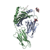
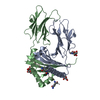
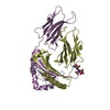
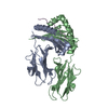
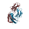
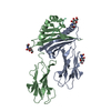
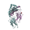
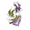
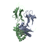
- PDB-1lnu: CRYSTAL STRUCTURE OF CLASS II MHC MOLECULE IAb BOUND TO EALPHA3K ... -
+ Open data

- Basic information
| Entry | Database: PDB / ID: 1lnu | |||||||||
|---|---|---|---|---|---|---|---|---|---|---|
| Title | CRYSTAL STRUCTURE OF CLASS II MHC MOLECULE IAb BOUND TO EALPHA3K PEPTIDE | |||||||||
 Components Components |
| |||||||||
 Keywords Keywords | SUGAR BINDING PROTEIN / Protein-peptide complex / T cell receptor / antigen presentation | |||||||||
| Function / homology |  Function and homology information Function and homology informationpositive regulation of antigen processing and presentation / positive regulation of alpha-beta T cell activation / positive regulation of T-helper 1 type immune response / B cell affinity maturation / protein antigen binding / positive regulation of T cell differentiation / toxic substance binding / multivesicular body / response to type II interferon / cellular response to type II interferon ...positive regulation of antigen processing and presentation / positive regulation of alpha-beta T cell activation / positive regulation of T-helper 1 type immune response / B cell affinity maturation / protein antigen binding / positive regulation of T cell differentiation / toxic substance binding / multivesicular body / response to type II interferon / cellular response to type II interferon / MHC class II protein complex / antigen processing and presentation of exogenous peptide antigen via MHC class II / peptide antigen binding / adaptive immune response / early endosome / lysosome / external side of plasma membrane / ubiquitin protein ligase binding / Golgi apparatus / cell surface Similarity search - Function | |||||||||
| Biological species |  | |||||||||
| Method |  X-RAY DIFFRACTION / SYNCHROTRON / MOLECULAR REPLACEMENT / Resolution: 2.5 Å X-RAY DIFFRACTION / SYNCHROTRON / MOLECULAR REPLACEMENT / Resolution: 2.5 Å | |||||||||
 Authors Authors | Liu, X. / Dai, S. / Crawford, F. / Fruge, R. / Marrack, P. / Kappler, J. | |||||||||
 Citation Citation | Journal: Proc.Natl.Acad.Sci.USA / Year: 2002 Title: Alternate interactions define the binding of peptides to the MHC molecule IA(b). Authors: Liu, X. / Dai, S. / Crawford, F. / Fruge, R. / Marrack, P. / Kappler, J. | |||||||||
| History |
| |||||||||
| Remark 999 | SEQUENCE Author states two separate sequences, an Ealpha3K peptide(1-13) and linker(14-28), which ...SEQUENCE Author states two separate sequences, an Ealpha3K peptide(1-13) and linker(14-28), which start from residue 1 to residue 28 were added to the N-terminus of the beta chain of the class II MHC IAB of chains B,D,F and H. |
- Structure visualization
| Structure viewer | Molecule: MolmilJmol/JSmol |
|---|
- Downloads & links
-Download
| PDBx/mmCIF format | 1lnu.cif.gz | 327.9 KB | Display | PDBx/mmCIF format |
|---|---|---|---|---|
| PDB format | pdb1lnu.ent.gz | 267.6 KB | Display | PDB format |
| PDBx/mmJSON format | 1lnu.json.gz | Tree view | PDBx/mmJSON format | |
| Others |  Other downloads Other downloads |
-Validation report
| Arichive directory | https://data.pdbj.org/pub/pdb/validation_reports/ln/1lnuftp://data.pdbj.org/pub/pdb/validation_reports/ln/1lnu | HTTPS FTP |
|---|
-Related structure data
| Related structure data |  2iadS S: Starting model for refinement |
|---|---|
| Similar structure data |
-Links
 PDBj
PDBj


- Assembly
| Deposited unit | 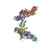
| ||||||||
|---|---|---|---|---|---|---|---|---|---|
| 1 | 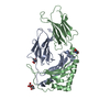
| ||||||||
| 2 | 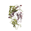
| ||||||||
| 3 | 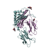
| ||||||||
| 4 | 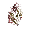
| ||||||||
| Unit cell |
| ||||||||
| Details | One molecule includes one alpha chain and one beta chain with the peptide |
-Components
| #1: Protein | Mass: 20597.957 Da / Num. of mol.: 4 Source method: isolated from a genetically manipulated source Source: (gene. exp.)   Spodoptera frugiperda (fall armyworm) / References: UniProt: P14434 Spodoptera frugiperda (fall armyworm) / References: UniProt: P14434#2: Protein | Mass: 24938.830 Da / Num. of mol.: 4 Source method: isolated from a genetically manipulated source Source: (gene. exp.)  #3: Sugar | ChemComp-NAG /   Type: D-saccharide, beta linking / Mass: 221.208 Da / Num. of mol.: 12 Type: D-saccharide, beta linking / Mass: 221.208 Da / Num. of mol.: 12Source method: isolated from a genetically manipulated source Formula: C8H15NO6 #4: Water | ChemComp-HOH / |  Mass: 18.015 Da / Num. of mol.: 237 / Source method: isolated from a natural source / Formula: H2O Mass: 18.015 Da / Num. of mol.: 237 / Source method: isolated from a natural source / Formula: H2OHas protein modification | Y | |
|---|
-Experimental details
-Experiment
| Experiment | Method: X-RAY DIFFRACTION / Number of used crystals: 1 |
|---|
- Sample preparation
| Crystal | Density Matthews: 2.97 Å3/Da / Density % sol: 58.53 % | |||||||||||||||||||||||||||||||||||
|---|---|---|---|---|---|---|---|---|---|---|---|---|---|---|---|---|---|---|---|---|---|---|---|---|---|---|---|---|---|---|---|---|---|---|---|---|
| Crystal grow | Temperature: 298 K / Method: vapor diffusion, hanging drop / pH: 6 Details: PEG8000, MES, KH2PO4, pH 6.0, VAPOR DIFFUSION, HANGING DROP, temperature 298K | |||||||||||||||||||||||||||||||||||
| Crystal grow | *PLUS | |||||||||||||||||||||||||||||||||||
| Components of the solutions | *PLUS
|
-Data collection
| Diffraction | Mean temperature: 100 K |
|---|---|
| Diffraction source | Source: SYNCHROTRON / Site: APS  / Beamline: 19-BM / Wavelength: 0.99187 Å / Beamline: 19-BM / Wavelength: 0.99187 Å |
| Detector | Type: SDMS / Detector: AREA DETECTOR / Date: Dec 6, 2001 |
| Radiation | Monochromator: Yale mirrors / Protocol: SINGLE WAVELENGTH / Monochromatic (M) / Laue (L): M / Scattering type: x-ray |
| Radiation wavelength | Wavelength: 0.99187 Å / Relative weight: 1 |
| Reflection | Resolution: 2.5→40 Å / Num. all: 54497 / Num. obs: 54497 / % possible obs: 74.8 % / Observed criterion σ(F): 2 / Observed criterion σ(I): -3 / Redundancy: 2.2 % / Biso Wilson estimate: 14.4 Å2 / Rmerge(I) obs: 0.089 |
| Reflection shell | Resolution: 2.5→2.59 Å / Rmerge(I) obs: 0.352 / Num. unique all: 1829 / % possible all: 21.3 |
| Reflection | *PLUS Highest resolution: 2.5 Å / Lowest resolution: 40 Å |
| Reflection shell | *PLUS % possible obs: 21.3 % / Num. unique obs: 1829 / Mean I/σ(I) obs: 4.3 |
- Processing
| Software |
| |||||||||||||||||||||||||
|---|---|---|---|---|---|---|---|---|---|---|---|---|---|---|---|---|---|---|---|---|---|---|---|---|---|---|
| Refinement | Method to determine structure: MOLECULAR REPLACEMENT Starting model: PDB ENTRY 2IAD Resolution: 2.5→38.39 Å / Rfactor Rfree error: 0.006 / Data cutoff high absF: 53511.21 / Data cutoff high rms absF: 53511.21 / Data cutoff low absF: 0 / Isotropic thermal model: RESTRAINED / Cross valid method: THROUGHOUT / σ(F): 0 / σ(I): 0 / Stereochemistry target values: Engh & Huber
| |||||||||||||||||||||||||
| Solvent computation | Solvent model: FLAT MODEL / Bsol: 28.563 Å2 / ksol: 0.342183 e/Å3 | |||||||||||||||||||||||||
| Displacement parameters | Biso mean: 18.6 Å2 | |||||||||||||||||||||||||
| Refine analyze |
| |||||||||||||||||||||||||
| Refinement step | Cycle: LAST / Resolution: 2.5→38.39 Å
| |||||||||||||||||||||||||
| Refine LS restraints |
| |||||||||||||||||||||||||
| Refine LS restraints NCS | NCS model details: CONSTR | |||||||||||||||||||||||||
| LS refinement shell | Resolution: 2.5→2.66 Å / Rfactor Rfree error: 0.032 / Total num. of bins used: 6
| |||||||||||||||||||||||||
| Xplor file |
| |||||||||||||||||||||||||
| Refinement | *PLUS Highest resolution: 2.5 Å / Lowest resolution: 40 Å / Num. reflection obs: 54427 | |||||||||||||||||||||||||
| Solvent computation | *PLUS | |||||||||||||||||||||||||
| Displacement parameters | *PLUS | |||||||||||||||||||||||||
| Refine LS restraints | *PLUS
| |||||||||||||||||||||||||
| LS refinement shell | *PLUS Lowest resolution: 2.59 Å / Rfactor Rfree: 0.313 / Num. reflection Rfree: 66 / Rfactor Rwork: 0.277 / Num. reflection obs: 1591 |
