 Movie
Movie Controller

[English] 日本語
 Yorodumi
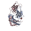
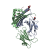
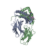
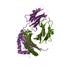
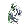
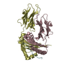
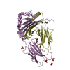
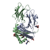
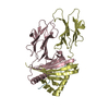
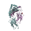
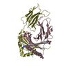
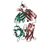
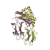
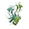
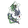
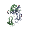
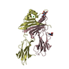
- PDB-1aqd: HLA-DR1 (DRA, DRB1 0101) HUMAN CLASS II HISTOCOMPATIBILITY PROTEI... -
+ Open data

- Basic information
| Entry | Database: PDB / ID: 1aqd | ||||||
|---|---|---|---|---|---|---|---|
| Title | HLA-DR1 (DRA, DRB1 0101) HUMAN CLASS II HISTOCOMPATIBILITY PROTEIN (EXTRACELLULAR DOMAIN) COMPLEXED WITH ENDOGENOUS PEPTIDE | ||||||
 Components Components |
| ||||||
 Keywords Keywords | COMPLEX (MHC PROTEIN/ANTIGEN) / COMPLEX (MHC PROTEIN-ANTIGEN) / HISTOCOMPATIBILITY ANTIGEN / COMPLEX (MHC PROTEIN-ANTIGEN) complex | ||||||
| Function / homology |  Function and homology information Function and homology informationregulation of interleukin-4 production / regulation of interleukin-10 production / autolysosome membrane / myeloid dendritic cell antigen processing and presentation / antigen processing and presentation of endogenous peptide antigen via MHC class II / regulation of T-helper cell differentiation / positive regulation of CD4-positive, CD25-positive, alpha-beta regulatory T cell differentiation / positive regulation of CD4-positive, alpha-beta T cell activation / antigen processing and presentation of peptide or polysaccharide antigen via MHC class II / positive regulation of T cell mediated immune response to tumor cell ...regulation of interleukin-4 production / regulation of interleukin-10 production / autolysosome membrane / myeloid dendritic cell antigen processing and presentation / antigen processing and presentation of endogenous peptide antigen via MHC class II / regulation of T-helper cell differentiation / positive regulation of CD4-positive, CD25-positive, alpha-beta regulatory T cell differentiation / positive regulation of CD4-positive, alpha-beta T cell activation / antigen processing and presentation of peptide or polysaccharide antigen via MHC class II / positive regulation of T cell mediated immune response to tumor cell / positive regulation of memory T cell differentiation / positive regulation of monocyte differentiation / inflammatory response to antigenic stimulus / CD4 receptor binding / positive regulation of memory T cell activation / T cell mediated cytotoxicity directed against tumor cell target / positive regulation of CD8-positive, alpha-beta T cell activation / CD8-positive, alpha-beta T cell activation / T-helper 1 type immune response / positive regulation of CD8-positive, alpha-beta T cell proliferation / intermediate filament / transport vesicle membrane / antigen processing and presentation of endogenous peptide antigen via MHC class I via ER pathway, TAP-dependent / TAP complex binding / antigen processing and presentation of exogenous peptide antigen via MHC class I / Golgi medial cisterna / Translocation of ZAP-70 to Immunological synapse / Phosphorylation of CD3 and TCR zeta chains / polysaccharide binding / CD8 receptor binding / negative regulation of type II interferon production / protection from natural killer cell mediated cytotoxicity / humoral immune response / beta-2-microglobulin binding / Generation of second messenger molecules / macrophage differentiation / endoplasmic reticulum exit site / immunological synapse / Co-inhibition by PD-1 / TAP binding / epidermis development / detection of bacterium / antigen processing and presentation of endogenous peptide antigen via MHC class Ib / antigen processing and presentation of endogenous peptide antigen via MHC class I via ER pathway, TAP-independent / negative regulation of T cell proliferation / T cell receptor binding / MHC class II antigen presentation / Nef mediated downregulation of MHC class I complex cell surface expression / positive regulation of insulin secretion involved in cellular response to glucose stimulus / trans-Golgi network membrane / Endosomal/Vacuolar pathway / T cell mediated cytotoxicity / Antigen Presentation: Folding, assembly and peptide loading of class I MHC / lumenal side of endoplasmic reticulum membrane / protein tetramerization / peptide antigen assembly with MHC class I protein complex / ER to Golgi transport vesicle membrane / negative regulation of inflammatory response to antigenic stimulus / clathrin-coated endocytic vesicle membrane / MHC class I peptide loading complex / positive regulation of T cell cytokine production / antigen processing and presentation of endogenous peptide antigen via MHC class I / MHC class I protein complex / peptide antigen assembly with MHC class II protein complex / MHC class II protein complex / positive regulation of T cell mediated cytotoxicity / structural constituent of cytoskeleton / antigen processing and presentation of exogenous peptide antigen via MHC class II / positive regulation of immune response / peptide antigen binding / cognition / positive regulation of type II interferon production / phagocytic vesicle membrane / recycling endosome membrane / positive regulation of T cell activation / Interferon gamma signaling / Immunoregulatory interactions between a Lymphoid and a non-Lymphoid cell / Interferon alpha/beta signaling / endocytic vesicle membrane / MHC class II protein complex binding / Downstream TCR signaling / late endosome membrane / T cell receptor signaling pathway / antibacterial humoral response / E3 ubiquitin ligases ubiquitinate target proteins / ER-Phagosome pathway / early endosome membrane / adaptive immune response / lysosome / defense response to Gram-positive bacterium / immune response / signaling receptor binding / Golgi membrane / external side of plasma membrane / innate immune response / lysosomal membrane / endoplasmic reticulum membrane / SARS-CoV-2 activates/modulates innate and adaptive immune responses / cell surface / endoplasmic reticulum Similarity search - Function | ||||||
| Biological species |  Homo sapiens (human) Homo sapiens (human) | ||||||
| Method |  X-RAY DIFFRACTION / SYNCHROTRON / MOLECULAR REPLACEMENT / Resolution: 2.45 Å X-RAY DIFFRACTION / SYNCHROTRON / MOLECULAR REPLACEMENT / Resolution: 2.45 Å | ||||||
 Authors Authors | Murthy, V.L. / Stern, L.J. | ||||||
 Citation Citation | Journal: Structure / Year: 1997 Title: The class II MHC protein HLA-DR1 in complex with an endogenous peptide: implications for the structural basis of the specificity of peptide binding. Authors: Murthy, V.L. / Stern, L.J. #1: Journal: Thesis / Year: 1996Title: Three Dimensional Structure of a Human Class II Mhc Protein Hla-Dr1 Bound to an Endogenous Peptide Authors: Murthy, V.L. #2: Journal: Nature / Year: 1994Title: Crystal Structure of the Human Class II Mhc Protein Hla-Dr1 Complexed with an Influenza Virus Peptide Authors: Stern, L.J. / Brown, J.H. / Jardetzky, T.S. / Gorga, J.C. / Urban, R.G. / Strominger, J.L. / Wiley, D.C. #3: Journal: Nature / Year: 1993Title: Three-Dimensional Structure of the Human Class II Histocompatibility Antigen Hla-Dr1 Authors: Brown, J.H. / Jardetzky, T.S. / Gorga, J.C. / Stern, L.J. / Urban, R.G. / Strominger, J.L. / Wiley, D.C. | ||||||
| History |
|
- Structure visualization
| Structure viewer | Molecule: MolmilJmol/JSmol |
|---|
- Downloads & links
-Download
| PDBx/mmCIF format | 1aqd.cif.gz | 319.3 KB | Display | PDBx/mmCIF format |
|---|---|---|---|---|
| PDB format | pdb1aqd.ent.gz | 258.6 KB | Display | PDB format |
| PDBx/mmJSON format | 1aqd.json.gz | Tree view | PDBx/mmJSON format | |
| Others |  Other downloads Other downloads |
-Validation report
| Arichive directory | https://data.pdbj.org/pub/pdb/validation_reports/aq/1aqdftp://data.pdbj.org/pub/pdb/validation_reports/aq/1aqd | HTTPS FTP |
|---|
-Related structure data
| Related structure data | 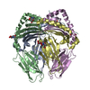 1dlhS S: Starting model for refinement |
|---|---|
| Similar structure data |

-Links
 PDBj
PDBj


- Assembly
| Deposited unit | 
| ||||||||||||||||||||||||||||
|---|---|---|---|---|---|---|---|---|---|---|---|---|---|---|---|---|---|---|---|---|---|---|---|---|---|---|---|---|---|
| 1 | 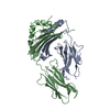
| ||||||||||||||||||||||||||||
| 2 | 
| ||||||||||||||||||||||||||||
| 3 | 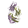
| ||||||||||||||||||||||||||||
| 4 | 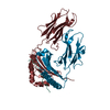
| ||||||||||||||||||||||||||||
| Unit cell |
| ||||||||||||||||||||||||||||
| Noncrystallographic symmetry (NCS) | NCS domain:
NCS oper:
| ||||||||||||||||||||||||||||
| Details | THERE ARE FOUR MOLECULES IN THIS COORDINATE SET EACH MOLECULE HAS ONE ALPHA CHAIN : RESIDUES 3-182 EACH MOLECULE HAS ONE BETA CHAIN : RESIDUES 4-190 EACH MOLECULE HAS ONE PEPTIDE CHAIN: RESIDUES 3-14 MOLECULE THREE IS THE MOST WELL DEFINED. MOLECULES 1 - 2 AND 3 - 4 EACH FORM THE SO-CALLED "DIMER OF DIMERS". |
-Components
| #1: Protein | Mass: 22222.027 Da / Num. of mol.: 4 / Fragment: SECRETED EXTRACELLULAR DOMAINS Source method: isolated from a genetically manipulated source Source: (gene. exp.) Homo sapiens (human) / Tissue: LYMPHOID / Cellular location: PLASMA MEMBRANE / Gene: DRA*0101, DRB1*0101 / Organ: PLASMA / Cell line (production host): SF9 / Production host:   Spodoptera frugiperda (fall armyworm) / References: UniProt: P01903 Spodoptera frugiperda (fall armyworm) / References: UniProt: P01903#2: Protein | Mass: 22956.596 Da / Num. of mol.: 4 / Fragment: SECRETED EXTRACELLULAR DOMAINS Source method: isolated from a genetically manipulated source Source: (gene. exp.) Homo sapiens (human) / Tissue: LYMPHOID / Cellular location: PLASMA MEMBRANE / Gene: DRA*0101, DRB1*0101 / Organ: PLASMA / Cell line (production host): SF9 / Production host: Spodoptera frugiperda (fall armyworm) / References: UniProt: P04229, UniProt: P01911*PLUS#3: Protein/peptide | Mass: 1858.045 Da / Num. of mol.: 4 / Fragment: ANTIGENIC PEPTIDE Source method: isolated from a genetically manipulated source Details: HLA-DR1 IS A CLASS II MHC PROTEIN, HLA-A2 IS A CLASS I HISTOCOMPATIBILITY ANTIGEN Source: (gene. exp.) Homo sapiens (human) / References: UniProt: P01892, UniProt: P04439*PLUS#4: Water | ChemComp-HOH / |  Mass: 18.015 Da / Num. of mol.: 152 / Source method: isolated from a natural source / Formula: H2O Mass: 18.015 Da / Num. of mol.: 152 / Source method: isolated from a natural source / Formula: H2OHas protein modification | Y | |
|---|
-Experimental details
-Experiment
| Experiment | Method: X-RAY DIFFRACTION / Number of used crystals: 1 |
|---|
- Sample preparation
| Crystal | Density Matthews: 3.04 Å3/Da / Density % sol: 56.2 % | ||||||||||||||||||||
|---|---|---|---|---|---|---|---|---|---|---|---|---|---|---|---|---|---|---|---|---|---|
| Crystal grow | pH: 3.5 Details: CRYSTALS GREW AS NEEDLES FROM 10MG/ML HLA-DR1 / PEPTIDE COMPLEX, 15% PEG 4000, 100MM GLYCINE, PH 3.5, AT ROOM TEMPERATURE, OVER ONE WEEK. Temp details: room temp | ||||||||||||||||||||
| Crystal | *PLUS | ||||||||||||||||||||
| Crystal grow | *PLUS Method: unknown | ||||||||||||||||||||
| Components of the solutions | *PLUS
|
-Data collection
| Diffraction | Mean temperature: 98 K |
|---|---|
| Diffraction source | Source: SYNCHROTRON / Site: CHESS  / Beamline: F1 / Wavelength: 0.918 / Beamline: F1 / Wavelength: 0.918 |
| Detector | Type: FUJI / Detector: IMAGE PLATE / Date: Feb 1, 1995 |
| Radiation | Monochromatic (M) / Laue (L): M / Scattering type: x-ray |
| Radiation wavelength | Wavelength: 0.918 Å / Relative weight: 1 |
| Reflection | Resolution: 2.45→20 Å / Num. obs: 73538 / % possible obs: 85 % / Observed criterion σ(I): 2 / Redundancy: 2.1 % / Biso Wilson estimate: 37.2 Å2 / Rsym value: 0.07 / Net I/σ(I): 14.9 |
| Reflection shell | Resolution: 2.45→2.57 Å / Redundancy: 1.8 % / Mean I/σ(I) obs: 4.4 / Rsym value: 0.331 / % possible all: 70 |
| Reflection | *PLUS Num. measured all: 157895 / Rmerge(I) obs: 0.07 |
| Reflection shell | *PLUS % possible obs: 70 % / Num. unique obs: 7700 / Num. measured obs: 13661 / Rmerge(I) obs: 0.331 |
- Processing
| Software |
| ||||||||||||||||||||||||||||||||||||||||||||||||||||||||||||
|---|---|---|---|---|---|---|---|---|---|---|---|---|---|---|---|---|---|---|---|---|---|---|---|---|---|---|---|---|---|---|---|---|---|---|---|---|---|---|---|---|---|---|---|---|---|---|---|---|---|---|---|---|---|---|---|---|---|---|---|---|---|
| Refinement | Method to determine structure: MOLECULAR REPLACEMENT Starting model: PDB ENTRY 1DLH Resolution: 2.45→6 Å / Isotropic thermal model: RESTRAINED / Cross valid method: THROUGHOUT / σ(F): 2 / Details: REFMAC USED IN LATER STAGES OF REFINEMENT.
| ||||||||||||||||||||||||||||||||||||||||||||||||||||||||||||
| Displacement parameters | Biso mean: 40.8 Å2 | ||||||||||||||||||||||||||||||||||||||||||||||||||||||||||||
| Refine analyze |
| ||||||||||||||||||||||||||||||||||||||||||||||||||||||||||||
| Refinement step | Cycle: LAST / Resolution: 2.45→6 Å
| ||||||||||||||||||||||||||||||||||||||||||||||||||||||||||||
| Refine LS restraints |
| ||||||||||||||||||||||||||||||||||||||||||||||||||||||||||||
| Refine LS restraints NCS |
| ||||||||||||||||||||||||||||||||||||||||||||||||||||||||||||
| LS refinement shell | Resolution: 2.45→2.57 Å / Total num. of bins used: 8
| ||||||||||||||||||||||||||||||||||||||||||||||||||||||||||||
| Xplor file |
| ||||||||||||||||||||||||||||||||||||||||||||||||||||||||||||
| Software | *PLUS Name: X-PLOR / Version: 3.1 / Classification: refinement | ||||||||||||||||||||||||||||||||||||||||||||||||||||||||||||
| Refine LS restraints | *PLUS
|
