 Movie
Movie Controller


+ Open data

- Basic information
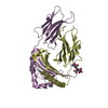
| Entry | Database: PDB / ID: 5v4n | ||||||
|---|---|---|---|---|---|---|---|
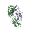
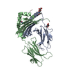
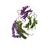
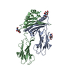
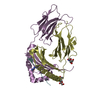
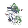
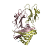
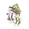
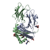
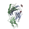
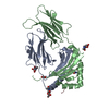
| Title | Structure of HLA-DR1 with bound alpha3(135-145) peptide | ||||||
 Components Components |
| ||||||
 Keywords Keywords | IMMUNE SYSTEM / HLA-DR / immune complex / antigen presenation | ||||||
| Function / homology |  Function and homology information Function and homology informationregulation of interleukin-4 production / regulation of interleukin-10 production / autolysosome membrane / myeloid dendritic cell antigen processing and presentation / antigen processing and presentation of endogenous peptide antigen via MHC class II / regulation of T-helper cell differentiation / positive regulation of CD4-positive, CD25-positive, alpha-beta regulatory T cell differentiation / positive regulation of CD4-positive, alpha-beta T cell activation / antigen processing and presentation of peptide or polysaccharide antigen via MHC class II / positive regulation of T cell mediated immune response to tumor cell ...regulation of interleukin-4 production / regulation of interleukin-10 production / autolysosome membrane / myeloid dendritic cell antigen processing and presentation / antigen processing and presentation of endogenous peptide antigen via MHC class II / regulation of T-helper cell differentiation / positive regulation of CD4-positive, CD25-positive, alpha-beta regulatory T cell differentiation / positive regulation of CD4-positive, alpha-beta T cell activation / antigen processing and presentation of peptide or polysaccharide antigen via MHC class II / positive regulation of T cell mediated immune response to tumor cell / positive regulation of memory T cell differentiation / positive regulation of monocyte differentiation / inflammatory response to antigenic stimulus / CD4 receptor binding / T-helper 1 type immune response / intermediate filament / transport vesicle membrane / Translocation of ZAP-70 to Immunological synapse / Phosphorylation of CD3 and TCR zeta chains / polysaccharide binding / negative regulation of type II interferon production / humoral immune response / Generation of second messenger molecules / macrophage differentiation / immunological synapse / Co-inhibition by PD-1 / epidermis development / detection of bacterium / negative regulation of T cell proliferation / T cell receptor binding / MHC class II antigen presentation / positive regulation of insulin secretion involved in cellular response to glucose stimulus / trans-Golgi network membrane / lumenal side of endoplasmic reticulum membrane / protein tetramerization / ER to Golgi transport vesicle membrane / negative regulation of inflammatory response to antigenic stimulus / clathrin-coated endocytic vesicle membrane / peptide antigen assembly with MHC class II protein complex / MHC class II protein complex / positive regulation of T cell mediated cytotoxicity / structural constituent of cytoskeleton / antigen processing and presentation of exogenous peptide antigen via MHC class II / positive regulation of immune response / peptide antigen binding / cognition / positive regulation of T cell activation / Interferon gamma signaling / endocytic vesicle membrane / MHC class II protein complex binding / Downstream TCR signaling / late endosome membrane / T cell receptor signaling pathway / early endosome membrane / adaptive immune response / lysosome / immune response / Golgi membrane / external side of plasma membrane / lysosomal membrane / cell surface / signal transduction / : / extracellular exosome / membrane / plasma membrane Similarity search - Function | ||||||
| Biological species |  Homo sapiens (human) Homo sapiens (human) | ||||||
| Method |  X-RAY DIFFRACTION / SYNCHROTRON / MOLECULAR REPLACEMENT / Resolution: 3.405 Å X-RAY DIFFRACTION / SYNCHROTRON / MOLECULAR REPLACEMENT / Resolution: 3.405 Å | ||||||
 Authors Authors | Petersen, J. / Rossjohn, J. | ||||||
 Citation Citation | Journal: Nature / Year: 2017 Title: Dominant protection from HLA-linked autoimmunity by antigen-specific regulatory T cells. Authors: Ooi, J.D. / Petersen, J. / Tan, Y.H. / Huynh, M. / Willett, Z.J. / Ramarathinam, S.H. / Eggenhuizen, P.J. / Loh, K.L. / Watson, K.A. / Gan, P.Y. / Alikhan, M.A. / Dudek, N.L. / Handel, A. / ...Authors: Ooi, J.D. / Petersen, J. / Tan, Y.H. / Huynh, M. / Willett, Z.J. / Ramarathinam, S.H. / Eggenhuizen, P.J. / Loh, K.L. / Watson, K.A. / Gan, P.Y. / Alikhan, M.A. / Dudek, N.L. / Handel, A. / Hudson, B.G. / Fugger, L. / Power, D.A. / Holt, S.G. / Coates, P.T. / Gregersen, J.W. / Purcell, A.W. / Holdsworth, S.R. / La Gruta, N.L. / Reid, H.H. / Rossjohn, J. / Kitching, A.R. | ||||||
| History |
|
- Structure visualization
| Structure viewer | Molecule: MolmilJmol/JSmol |
|---|
- Downloads & links
-Download
| PDBx/mmCIF format | 5v4n.cif.gz | 324.4 KB | Display | PDBx/mmCIF format |
|---|---|---|---|---|
| PDB format | pdb5v4n.ent.gz | 264.8 KB | Display | PDB format |
| PDBx/mmJSON format | 5v4n.json.gz | Tree view | PDBx/mmJSON format | |
| Others |  Other downloads Other downloads |
-Validation report
| Arichive directory | https://data.pdbj.org/pub/pdb/validation_reports/v4/5v4nftp://data.pdbj.org/pub/pdb/validation_reports/v4/5v4n | HTTPS FTP |
|---|
-Related structure data
| Related structure data |  5v4mC 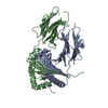 3qxaS S: Starting model for refinement C: citing same article ( |
|---|---|
| Similar structure data |
-Links
 PDBj
PDBj


- Assembly
| Deposited unit | 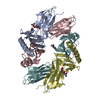
| ||||||||
|---|---|---|---|---|---|---|---|---|---|
| 1 | 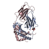
| ||||||||
| 2 | 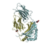
| ||||||||
| Unit cell |
|
-Components
| #1: Protein | Mass: 21919.594 Da / Num. of mol.: 2 Source method: isolated from a genetically manipulated source Source: (gene. exp.) Homo sapiens (human) / Gene: HLA-DRA, HLA-DRA1 / Production host:  Trichoplusia ni (cabbage looper) / References: UniProt: P01903 Trichoplusia ni (cabbage looper) / References: UniProt: P01903#2: Protein | Mass: 24609.412 Da / Num. of mol.: 2 Source method: isolated from a genetically manipulated source Source: (gene. exp.) Homo sapiens (human) / Gene: HLA-DRB1 / Production host: Trichoplusia ni (cabbage looper) / References: UniProt: P04229, UniProt: P01911*PLUS#3: Sugar |   Type: D-saccharide, beta linking / Mass: 221.208 Da / Num. of mol.: 3 Type: D-saccharide, beta linking / Mass: 221.208 Da / Num. of mol.: 3Source method: isolated from a genetically manipulated source Formula: C8H15NO6 Has protein modification | Y | |
|---|
-Experimental details
-Experiment
| Experiment | Method: X-RAY DIFFRACTION / Number of used crystals: 1 |
|---|
- Sample preparation
| Crystal | Density Matthews: 3.16 Å3/Da / Density % sol: 61.03 % |
|---|---|
| Crystal grow | Temperature: 298.15 K / Method: vapor diffusion, hanging drop / pH: 7 Details: 23% PEG 3350, 0.1M KNO3, and 0.1M bis-tris-propane (pH 7.0) |
-Data collection
| Diffraction | Mean temperature: 100 K |
|---|---|
| Diffraction source | Source: SYNCHROTRON / Site: Australian Synchrotron  / Beamline: MX2 / Wavelength: 0.9537 Å / Beamline: MX2 / Wavelength: 0.9537 Å |
| Detector | Type: ADSC QUANTUM 315r / Detector: CCD / Date: Jun 10, 2016 |
| Radiation | Protocol: SINGLE WAVELENGTH / Monochromatic (M) / Laue (L): M / Scattering type: x-ray |
| Radiation wavelength | Wavelength: 0.9537 Å / Relative weight: 1 |
| Reflection | Resolution: 3.405→53.831 Å / Num. obs: 16628 / % possible obs: 99 % / Redundancy: 2 % / CC1/2: 0.942 / Rmerge(I) obs: 0.1099 / Net I/σ(I): 6.04 |
| Reflection shell | Redundancy: 2 % / Rmerge(I) obs: 0.3648 / Mean I/σ(I) obs: 2.6 / Num. unique obs: 1610 / CC1/2: 0.651 / % possible all: 99 |
- Processing
| Software |
| |||||||||||||||||||||||||||||||||||||||||||||||||
|---|---|---|---|---|---|---|---|---|---|---|---|---|---|---|---|---|---|---|---|---|---|---|---|---|---|---|---|---|---|---|---|---|---|---|---|---|---|---|---|---|---|---|---|---|---|---|---|---|---|---|
| Refinement | Method to determine structure: MOLECULAR REPLACEMENT Starting model: 3QXA Resolution: 3.405→53.831 Å / SU ML: 0.4 / Cross valid method: THROUGHOUT / σ(F): 1.35 / Phase error: 26.08
| |||||||||||||||||||||||||||||||||||||||||||||||||
| Solvent computation | Shrinkage radii: 0.9 Å / VDW probe radii: 1.11 Å | |||||||||||||||||||||||||||||||||||||||||||||||||
| Refinement step | Cycle: LAST / Resolution: 3.405→53.831 Å
| |||||||||||||||||||||||||||||||||||||||||||||||||
| Refine LS restraints |
| |||||||||||||||||||||||||||||||||||||||||||||||||
| LS refinement shell |
| |||||||||||||||||||||||||||||||||||||||||||||||||
| Refinement TLS params. | Method: refined / Origin x: 13.411 Å / Origin y: 109.1652 Å / Origin z: 136.7139 Å
| |||||||||||||||||||||||||||||||||||||||||||||||||
| Refinement TLS group | Selection details: all |
