 Movie
Movie Controller

[English] 日本語
 Yorodumi
- PDB-7rjj: Crystal Structure of the Peptidoglycan Binding Domain of the Oute... -
+ Open data

- Basic information
| Entry | Database: PDB / ID: 7rjj | ||||||
|---|---|---|---|---|---|---|---|
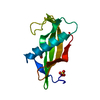
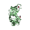
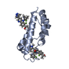
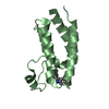
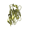
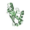
| Title | Crystal Structure of the Peptidoglycan Binding Domain of the Outer Membrane Protein (OmpA) from Klebsiella pneumoniae with bound D-alanine | ||||||
 Components Components | OmpA family protein | ||||||
 Keywords Keywords | PEPTIDE BINDING PROTEIN / Structural Genomics / Center for Structural Genomics of Infectious Diseases / CSGID / Outer Membrane Protein | ||||||
| Function / homology | Outer membrane protein, bacterial / : / OmpA-like domain profile. / OmpA family / OmpA-like domain / OmpA-like domain superfamily / membrane / D-ALANINE / OmpA family protein Function and homology information Function and homology information | ||||||
| Biological species |  Klebsiella pneumoniae subsp. pneumoniae (bacteria) Klebsiella pneumoniae subsp. pneumoniae (bacteria) | ||||||
| Method |  X-RAY DIFFRACTION / SYNCHROTRON / SAD / Resolution: 1.88 Å X-RAY DIFFRACTION / SYNCHROTRON / SAD / Resolution: 1.88 Å | ||||||
 Authors Authors | Minasov, G. / Shuvalova, L. / Kiryukhina, O. / Dubrovska, I. / Satchell, K.J.F. / Center for Structural Genomics of Infectious Diseases (CSGID) | ||||||
 Citation Citation | Journal: Microbiol Resour Announc / Year: 2023 Title: A Structural Systems Biology Approach to High-Risk CG23 Klebsiella pneumoniae. Authors: Inniss, N.L. / Kochan, T.J. / Minasov, G. / Wawrzak, Z. / Chang, C. / Tan, K. / Shuvalova, L. / Kiryukhina, O. / Pshenychnyi, S. / Wu, R. / Dubrovska, I. / Babnigg, G. / Endres, M. / ...Authors: Inniss, N.L. / Kochan, T.J. / Minasov, G. / Wawrzak, Z. / Chang, C. / Tan, K. / Shuvalova, L. / Kiryukhina, O. / Pshenychnyi, S. / Wu, R. / Dubrovska, I. / Babnigg, G. / Endres, M. / Anderson, W.F. / Hauser, A.R. / Joachimiak, A. / Satchell, K.J.F. | ||||||
| History |
|
- Structure visualization
| Structure viewer | Molecule: MolmilJmol/JSmol |
|---|
- Downloads & links
-Download
| PDBx/mmCIF format | 7rjj.cif.gz | 113.3 KB | Display | PDBx/mmCIF format |
|---|---|---|---|---|
| PDB format | pdb7rjj.ent.gz | 87 KB | Display | PDB format |
| PDBx/mmJSON format | 7rjj.json.gz | Tree view | PDBx/mmJSON format | |
| Others |  Other downloads Other downloads |
-Validation report
| Arichive directory | https://data.pdbj.org/pub/pdb/validation_reports/rj/7rjjftp://data.pdbj.org/pub/pdb/validation_reports/rj/7rjj | HTTPS FTP |
|---|
-Related structure data
| Related structure data |  6dt3C  6duxC  6dvvC  6dxnC  6e85C  6nauC  6nbgC  6ndiC  6wn5C  6wn8C 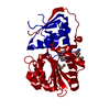 6x1lC  7tl5C 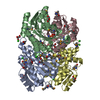 7tzpC C: citing same article ( |
|---|---|
| Similar structure data | |
| Other databases |


-Links
 PDBj
PDBj- Assembly
| Deposited unit | 
| ||||||||
|---|---|---|---|---|---|---|---|---|---|
| 1 | 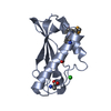
| ||||||||
| 2 | 
| ||||||||
| Unit cell |
|
-Components
| #1: Protein | Mass: 14108.505 Da / Num. of mol.: 2 Source method: isolated from a genetically manipulated source Source: (gene. exp.) Klebsiella pneumoniae subsp. pneumoniae (bacteria)Gene: C4Z38_016670, EJ893_12145, I5002_15200, JMY88_11465 / Plasmid: pMCSG53 / Production host: #2: Chemical |   Type: D-peptide linking / Mass: 89.093 Da / Num. of mol.: 2 / Source method: obtained synthetically / Formula: C3H7NO2 / Feature type: SUBJECT OF INVESTIGATION Type: D-peptide linking / Mass: 89.093 Da / Num. of mol.: 2 / Source method: obtained synthetically / Formula: C3H7NO2 / Feature type: SUBJECT OF INVESTIGATION#3: Chemical |   Mass: 35.453 Da / Num. of mol.: 2 / Source method: obtained synthetically / Formula: Cl Mass: 35.453 Da / Num. of mol.: 2 / Source method: obtained synthetically / Formula: Cl#4: Water | ChemComp-HOH / |  Mass: 18.015 Da / Num. of mol.: 177 / Source method: isolated from a natural source / Formula: H2O Mass: 18.015 Da / Num. of mol.: 177 / Source method: isolated from a natural source / Formula: H2OHas ligand of interest | Y | Has protein modification | Y | |
|---|
-Experimental details
-Experiment
| Experiment | Method: X-RAY DIFFRACTION / Number of used crystals: 1 |
|---|
- Sample preparation
| Crystal | Density Matthews: 2.37 Å3/Da / Density % sol: 48.2 % |
|---|---|
| Crystal grow | Temperature: 292 K / Method: vapor diffusion, sitting drop / pH: 6.5 Details: Protein: 9.4 mg/ml, 0.15M Sodium chloride, 0.01M Tris pH 8.3; Screen: PEG's II (C4), 0.2M Magnesium chloride, 0.1M MES pH 6.5, 10% (w/v) PEG4000; Cryo: 0.2M Magnesium chloride, 0.1M MES pH 6.5, 25% (w/v) PEG4000 |
-Data collection
| Diffraction | Mean temperature: 100 K / Serial crystal experiment: N |
|---|---|
| Diffraction source | Source: SYNCHROTRON / Site: APS  / Beamline: 21-ID-F / Wavelength: 0.97872 Å / Beamline: 21-ID-F / Wavelength: 0.97872 Å |
| Detector | Type: MARMOSAIC 300 mm CCD / Detector: CCD / Date: Feb 3, 2021 / Details: Be |
| Radiation | Monochromator: C(111) / Protocol: SINGLE WAVELENGTH / Monochromatic (M) / Laue (L): M / Scattering type: x-ray |
| Radiation wavelength | Wavelength: 0.97872 Å / Relative weight: 1 |
| Reflection | Resolution: 1.88→30 Å / Num. obs: 21507 / % possible obs: 99.9 % / Observed criterion σ(I): -3 / Redundancy: 11.4 % / Biso Wilson estimate: 17.3 Å2 / CC1/2: 0.988 / CC star: 0.997 / Rmerge(I) obs: 0.073 / Rpim(I) all: 0.023 / Rrim(I) all: 0.076 / Rsym value: 0.073 / Χ2: 0.837 / Net I/σ(I): 31.5 |
| Reflection shell | Resolution: 1.88→1.91 Å / Redundancy: 10.5 % / Rmerge(I) obs: 1.358 / Mean I/σ(I) obs: 1.8 / Num. unique obs: 1084 / CC1/2: 0.626 / CC star: 0.878 / Rpim(I) all: 0.439 / Rsym value: 1.358 / Χ2: 0.765 / % possible all: 100 |
- Processing
| Software |
| |||||||||||||||||||||||||||||||||||||||||||||||||||||||||||||||||||||||||||||||||||||||||||||||||||||||||||||||||||||||||||||||||||||||||||||||||||||||||||||||||||||||||||||||||||||||||||||||||||||||||||||||||||||||||||||||||
|---|---|---|---|---|---|---|---|---|---|---|---|---|---|---|---|---|---|---|---|---|---|---|---|---|---|---|---|---|---|---|---|---|---|---|---|---|---|---|---|---|---|---|---|---|---|---|---|---|---|---|---|---|---|---|---|---|---|---|---|---|---|---|---|---|---|---|---|---|---|---|---|---|---|---|---|---|---|---|---|---|---|---|---|---|---|---|---|---|---|---|---|---|---|---|---|---|---|---|---|---|---|---|---|---|---|---|---|---|---|---|---|---|---|---|---|---|---|---|---|---|---|---|---|---|---|---|---|---|---|---|---|---|---|---|---|---|---|---|---|---|---|---|---|---|---|---|---|---|---|---|---|---|---|---|---|---|---|---|---|---|---|---|---|---|---|---|---|---|---|---|---|---|---|---|---|---|---|---|---|---|---|---|---|---|---|---|---|---|---|---|---|---|---|---|---|---|---|---|---|---|---|---|---|---|---|---|---|---|---|---|---|---|---|---|---|---|---|---|---|---|---|---|---|---|---|---|
| Refinement | Method to determine structure: SAD / Resolution: 1.88→28.16 Å / Cor.coef. Fo:Fc: 0.938 / Cor.coef. Fo:Fc free: 0.924 / SU B: 7.379 / SU ML: 0.111 / Cross valid method: THROUGHOUT / σ(F): 0 / ESU R: 0.172 / ESU R Free: 0.147 / Stereochemistry target values: MAXIMUM LIKELIHOOD Details: HYDROGENS HAVE BEEN ADDED IN THE RIDING POSITIONS U VALUES : WITH TLS ADDED
| |||||||||||||||||||||||||||||||||||||||||||||||||||||||||||||||||||||||||||||||||||||||||||||||||||||||||||||||||||||||||||||||||||||||||||||||||||||||||||||||||||||||||||||||||||||||||||||||||||||||||||||||||||||||||||||||||
| Solvent computation | Ion probe radii: 0.8 Å / Shrinkage radii: 0.8 Å / VDW probe radii: 1.2 Å / Solvent model: MASK | |||||||||||||||||||||||||||||||||||||||||||||||||||||||||||||||||||||||||||||||||||||||||||||||||||||||||||||||||||||||||||||||||||||||||||||||||||||||||||||||||||||||||||||||||||||||||||||||||||||||||||||||||||||||||||||||||
| Displacement parameters | Biso max: 59.21 Å2 / Biso mean: 22.826 Å2 / Biso min: 6.07 Å2
| |||||||||||||||||||||||||||||||||||||||||||||||||||||||||||||||||||||||||||||||||||||||||||||||||||||||||||||||||||||||||||||||||||||||||||||||||||||||||||||||||||||||||||||||||||||||||||||||||||||||||||||||||||||||||||||||||
| Refinement step | Cycle: final / Resolution: 1.88→28.16 Å
| |||||||||||||||||||||||||||||||||||||||||||||||||||||||||||||||||||||||||||||||||||||||||||||||||||||||||||||||||||||||||||||||||||||||||||||||||||||||||||||||||||||||||||||||||||||||||||||||||||||||||||||||||||||||||||||||||
| Refine LS restraints |
| |||||||||||||||||||||||||||||||||||||||||||||||||||||||||||||||||||||||||||||||||||||||||||||||||||||||||||||||||||||||||||||||||||||||||||||||||||||||||||||||||||||||||||||||||||||||||||||||||||||||||||||||||||||||||||||||||
| LS refinement shell | Resolution: 1.882→1.93 Å / Rfactor Rfree error: 0 / Total num. of bins used: 20
| |||||||||||||||||||||||||||||||||||||||||||||||||||||||||||||||||||||||||||||||||||||||||||||||||||||||||||||||||||||||||||||||||||||||||||||||||||||||||||||||||||||||||||||||||||||||||||||||||||||||||||||||||||||||||||||||||
| Refinement TLS params. | Method: refined / Refine-ID: X-RAY DIFFRACTION
| |||||||||||||||||||||||||||||||||||||||||||||||||||||||||||||||||||||||||||||||||||||||||||||||||||||||||||||||||||||||||||||||||||||||||||||||||||||||||||||||||||||||||||||||||||||||||||||||||||||||||||||||||||||||||||||||||
| Refinement TLS group |
|
