 Movie
Movie Controller

[English] 日本語
 Yorodumi
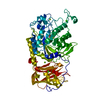
- PDB-6x1l: The crystal structure of a functional uncharacterized protein KP1... -
+ Open data

- Basic information
| Entry | Database: PDB / ID: 6x1l | ||||||
|---|---|---|---|---|---|---|---|
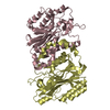
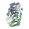
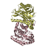
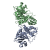
| Title | The crystal structure of a functional uncharacterized protein KP1_0663 from Klebsiella pneumoniae subsp. pneumoniae NTUH-K2044 | ||||||
 Components Components | WbbZ protein | ||||||
 Keywords Keywords | UNKNOWN FUNCTION / uncharacterized / Structural Genomics / Center for Structural Genomics of Infectious Diseases / CSGID | ||||||
| Function / homology | Polysaccharide pyruvyl transferase / Polysaccharide pyruvyl transferase / WbbZ protein Function and homology information Function and homology information | ||||||
| Biological species |  Klebsiella pneumoniae subsp. pneumoniae NTUH-K2044 (bacteria) Klebsiella pneumoniae subsp. pneumoniae NTUH-K2044 (bacteria) | ||||||
| Method |  X-RAY DIFFRACTION / SYNCHROTRON / SAD / Resolution: 2 Å X-RAY DIFFRACTION / SYNCHROTRON / SAD / Resolution: 2 Å | ||||||
 Authors Authors | Tan, K. / Wu, R. / Endres, M. / Joachimiak, A. / Center for Structural Genomics of Infectious Diseases (CSGID) | ||||||
| Funding support |  United States, 1items United States, 1items
| ||||||
 Citation Citation | Journal: Microbiol Resour Announc / Year: 2023 Title: A Structural Systems Biology Approach to High-Risk CG23 Klebsiella pneumoniae. Authors: Inniss, N.L. / Kochan, T.J. / Minasov, G. / Wawrzak, Z. / Chang, C. / Tan, K. / Shuvalova, L. / Kiryukhina, O. / Pshenychnyi, S. / Wu, R. / Dubrovska, I. / Babnigg, G. / Endres, M. / ...Authors: Inniss, N.L. / Kochan, T.J. / Minasov, G. / Wawrzak, Z. / Chang, C. / Tan, K. / Shuvalova, L. / Kiryukhina, O. / Pshenychnyi, S. / Wu, R. / Dubrovska, I. / Babnigg, G. / Endres, M. / Anderson, W.F. / Hauser, A.R. / Joachimiak, A. / Satchell, K.J.F. | ||||||
| History |
|
- Structure visualization
| Structure viewer | Molecule: MolmilJmol/JSmol |
|---|
- Downloads & links
-Download
| PDBx/mmCIF format | 6x1l.cif.gz | 145.3 KB | Display | PDBx/mmCIF format |
|---|---|---|---|---|
| PDB format | pdb6x1l.ent.gz | 95.6 KB | Display | PDB format |
| PDBx/mmJSON format | 6x1l.json.gz | Tree view | PDBx/mmJSON format | |
| Others |  Other downloads Other downloads |
-Validation report
| Arichive directory | https://data.pdbj.org/pub/pdb/validation_reports/x1/6x1lftp://data.pdbj.org/pub/pdb/validation_reports/x1/6x1l | HTTPS FTP |
|---|
-Related structure data
| Related structure data |  6dt3C  6duxC  6dvvC 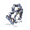 6dxnC 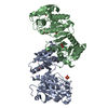 6e85C  6nauC 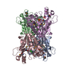 6nbgC 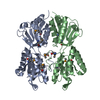 6ndiC  6wn5C  6wn8C  7rjjC  7tl5C  7tzpC C: citing same article ( |
|---|---|
| Similar structure data | |
| Other databases |


-Links
 PDBj
PDBj- Assembly
| Deposited unit | 
| ||||||||||||
|---|---|---|---|---|---|---|---|---|---|---|---|---|---|
| 1 | 
| ||||||||||||
| Unit cell |
|
-Components
| #1: Protein | Mass: 31878.037 Da / Num. of mol.: 1 Source method: isolated from a genetically manipulated source Source: (gene. exp.) Klebsiella pneumoniae subsp. pneumoniae NTUH-K2044 (bacteria)Gene: wbbZ / Plasmid: pMCSG73 / Production host: |
|---|---|
| #2: Water | ChemComp-HOH /  Mass: 18.015 Da / Num. of mol.: 32 / Source method: isolated from a natural source / Formula: H2O Mass: 18.015 Da / Num. of mol.: 32 / Source method: isolated from a natural source / Formula: H2O |
| Has ligand of interest | N |
| Has protein modification | Y |
-Experimental details
-Experiment
| Experiment | Method: X-RAY DIFFRACTION / Number of used crystals: 1 |
|---|
- Sample preparation
| Crystal | Density Matthews: 3.21 Å3/Da / Density % sol: 61.7 % |
|---|---|
| Crystal grow | Temperature: 297 K / Method: vapor diffusion, sitting drop / pH: 7 Details: 1.1 M sodium Malonate, 0.1 M HEPES:NaOH, 0.5 % (v/v) Jeffamine ED-2001 |
-Data collection
| Diffraction | Mean temperature: 100 K / Serial crystal experiment: N |
|---|---|
| Diffraction source | Source: SYNCHROTRON / Site: APS / Beamline: 19-ID / Wavelength: 0.97918 Å |
| Detector | Type: DECTRIS PILATUS3 S 6M / Detector: PIXEL / Date: Apr 24, 2018 |
| Radiation | Monochromator: Si 111 crystal / Protocol: SINGLE WAVELENGTH / Monochromatic (M) / Laue (L): M / Scattering type: x-ray |
| Radiation wavelength | Wavelength: 0.97918 Å / Relative weight: 1 |
| Reflection | Resolution: 2→41 Å / Num. obs: 27259 / % possible obs: 98.6 % / Observed criterion σ(I): -3 / Redundancy: 5.2 % / Biso Wilson estimate: 46.78 Å2 / CC1/2: 1 / CC star: 1 / Rmerge(I) obs: 0.074 / Rpim(I) all: 0.035 / Rrim(I) all: 0.082 / Χ2: 2.43 / Net I/σ(I): 38.4 |
| Reflection shell | Resolution: 2→2.03 Å / Redundancy: 4.5 % / Rmerge(I) obs: 0.685 / Mean I/σ(I) obs: 1.27 / Num. unique obs: 1163 / CC1/2: 0.85 / Rpim(I) all: 0.324 / Rrim(I) all: 0.761 / Χ2: 0.591 / % possible all: 85.6 |
- Processing
| Software |
| |||||||||||||||||||||||||||||||||||||||||||||||||||||||||||||||||||||||||||||||||||||||||||||||||||||||||||||||||||||||||||||
|---|---|---|---|---|---|---|---|---|---|---|---|---|---|---|---|---|---|---|---|---|---|---|---|---|---|---|---|---|---|---|---|---|---|---|---|---|---|---|---|---|---|---|---|---|---|---|---|---|---|---|---|---|---|---|---|---|---|---|---|---|---|---|---|---|---|---|---|---|---|---|---|---|---|---|---|---|---|---|---|---|---|---|---|---|---|---|---|---|---|---|---|---|---|---|---|---|---|---|---|---|---|---|---|---|---|---|---|---|---|---|---|---|---|---|---|---|---|---|---|---|---|---|---|---|---|---|
| Refinement | Method to determine structure: SAD / Resolution: 2→40.93 Å / SU ML: 0.2798 / Cross valid method: FREE R-VALUE / σ(F): 1.38 / Phase error: 28.6846 Stereochemistry target values: GeoStd + Monomer Library + CDL v1.2
| |||||||||||||||||||||||||||||||||||||||||||||||||||||||||||||||||||||||||||||||||||||||||||||||||||||||||||||||||||||||||||||
| Solvent computation | Shrinkage radii: 0.9 Å / VDW probe radii: 1.11 Å / Solvent model: FLAT BULK SOLVENT MODEL | |||||||||||||||||||||||||||||||||||||||||||||||||||||||||||||||||||||||||||||||||||||||||||||||||||||||||||||||||||||||||||||
| Displacement parameters | Biso mean: 65.48 Å2 | |||||||||||||||||||||||||||||||||||||||||||||||||||||||||||||||||||||||||||||||||||||||||||||||||||||||||||||||||||||||||||||
| Refinement step | Cycle: LAST / Resolution: 2→40.93 Å
| |||||||||||||||||||||||||||||||||||||||||||||||||||||||||||||||||||||||||||||||||||||||||||||||||||||||||||||||||||||||||||||
| Refine LS restraints |
| |||||||||||||||||||||||||||||||||||||||||||||||||||||||||||||||||||||||||||||||||||||||||||||||||||||||||||||||||||||||||||||
| LS refinement shell |
| |||||||||||||||||||||||||||||||||||||||||||||||||||||||||||||||||||||||||||||||||||||||||||||||||||||||||||||||||||||||||||||
| Refinement TLS params. | Method: refined / Refine-ID: X-RAY DIFFRACTION
| |||||||||||||||||||||||||||||||||||||||||||||||||||||||||||||||||||||||||||||||||||||||||||||||||||||||||||||||||||||||||||||
| Refinement TLS group |
|
