 Movie
Movie Controller

[English] 日本語
 Yorodumi
- PDB-6ndi: Crystal Structure of the Sugar Binding Domain of LacI Family Prot... -
+ Open data

- Basic information
| Entry | Database: PDB / ID: 6ndi | ||||||
|---|---|---|---|---|---|---|---|
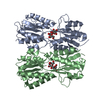
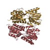
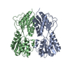
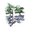
| Title | Crystal Structure of the Sugar Binding Domain of LacI Family Protein from Klebsiella pneumoniae | ||||||
 Components Components | Transcriptional regulator | ||||||
 Keywords Keywords | SUGAR BINDING PROTEIN / Structural Genomics / Center for Structural Genomics of Infectious Diseases / CSGID / Periplasmic Binding / Sugar Binding | ||||||
| Function / homology |  Function and homology information Function and homology information | ||||||
| Biological species |  Klebsiella pneumoniae subsp. pneumoniae (bacteria) Klebsiella pneumoniae subsp. pneumoniae (bacteria) | ||||||
| Method |  X-RAY DIFFRACTION / SYNCHROTRON / SAD / Resolution: 2.6 Å X-RAY DIFFRACTION / SYNCHROTRON / SAD / Resolution: 2.6 Å | ||||||
 Authors Authors | Minasov, G. / Shuvalova, L. / Wawrzak, Z. / Kiryukhina, O. / Dubrovska, I. / Anderson, W.F. / Satchell, K.J.F. / Joachimiak, A. / Center for Structural Genomics of Infectious Diseases (CSGID) | ||||||
 Citation Citation | Journal: Microbiol Resour Announc / Year: 2023 Title: A Structural Systems Biology Approach to High-Risk CG23 Klebsiella pneumoniae. Authors: Inniss, N.L. / Kochan, T.J. / Minasov, G. / Wawrzak, Z. / Chang, C. / Tan, K. / Shuvalova, L. / Kiryukhina, O. / Pshenychnyi, S. / Wu, R. / Dubrovska, I. / Babnigg, G. / Endres, M. / ...Authors: Inniss, N.L. / Kochan, T.J. / Minasov, G. / Wawrzak, Z. / Chang, C. / Tan, K. / Shuvalova, L. / Kiryukhina, O. / Pshenychnyi, S. / Wu, R. / Dubrovska, I. / Babnigg, G. / Endres, M. / Anderson, W.F. / Hauser, A.R. / Joachimiak, A. / Satchell, K.J.F. | ||||||
| History |
|
- Structure visualization
| Structure viewer | Molecule: MolmilJmol/JSmol |
|---|
- Downloads & links
-Download
| PDBx/mmCIF format | 6ndi.cif.gz | 228.5 KB | Display | PDBx/mmCIF format |
|---|---|---|---|---|
| PDB format | pdb6ndi.ent.gz | 183.7 KB | Display | PDB format |
| PDBx/mmJSON format | 6ndi.json.gz | Tree view | PDBx/mmJSON format | |
| Others |  Other downloads Other downloads |
-Validation report
| Arichive directory | https://data.pdbj.org/pub/pdb/validation_reports/nd/6ndiftp://data.pdbj.org/pub/pdb/validation_reports/nd/6ndi | HTTPS FTP |
|---|
-Related structure data
| Related structure data |  6dt3C  6duxC  6dvvC  6dxnC  6e85C  6nauC  6nbgC  6wn5C  6wn8C  6x1lC  7rjjC  7tl5C  7tzpC C: citing same article ( |
|---|---|
| Similar structure data | |
| Other databases |

-Links
 PDBj
PDBj


- Assembly
| Deposited unit | 
| ||||||||||||||||||
|---|---|---|---|---|---|---|---|---|---|---|---|---|---|---|---|---|---|---|---|
| 1 |
| ||||||||||||||||||
| Unit cell |
| ||||||||||||||||||
| Noncrystallographic symmetry (NCS) | NCS domain:
NCS domain segments: Component-ID: _ / Ens-ID: 1 / Beg auth comp-ID: PRO / Beg label comp-ID: PRO / End auth comp-ID: SER / End label comp-ID: SER / Refine code: _ / Auth seq-ID: 61 - 330 / Label seq-ID: 64 - 333
|
-Components
| #1: Protein | Mass: 36938.145 Da / Num. of mol.: 2 Source method: isolated from a genetically manipulated source Source: (gene. exp.) Klebsiella pneumoniae subsp. pneumoniae (bacteria)Gene: AN676_0312450 / Plasmid: pMCSG53 / Production host: #2: Water | ChemComp-HOH / |  Mass: 18.015 Da / Num. of mol.: 157 / Source method: isolated from a natural source / Formula: H2O Mass: 18.015 Da / Num. of mol.: 157 / Source method: isolated from a natural source / Formula: H2OHas protein modification | Y | |
|---|
-Experimental details
-Experiment
| Experiment | Method: X-RAY DIFFRACTION / Number of used crystals: 1 |
|---|
- Sample preparation
| Crystal | Density Matthews: 2.5 Å3/Da / Density % sol: 51 % |
|---|---|
| Crystal grow | Temperature: 292 K / Method: vapor diffusion, sitting drop / pH: 7 Details: Protein: 3.0 mg/ml, 0.01M Tris-HCl pH 8.3; Screen: Classics II (C1), 3.5M Sodium formate pH 7.0; Cryo: 4.0M Sodium formate. |
-Data collection
| Diffraction | Mean temperature: 100 K / Serial crystal experiment: N |
|---|---|
| Diffraction source | Source: SYNCHROTRON / Site: APS  / Beamline: 21-ID-G / Wavelength: 0.97856 Å / Beamline: 21-ID-G / Wavelength: 0.97856 Å |
| Detector | Type: MARMOSAIC 300 mm CCD / Detector: CCD / Date: Nov 15, 2018 / Details: C(111) |
| Radiation | Monochromator: Be / Protocol: SINGLE WAVELENGTH / Monochromatic (M) / Laue (L): M / Scattering type: x-ray |
| Radiation wavelength | Wavelength: 0.97856 Å / Relative weight: 1 |
| Reflection | Resolution: 2.6→30 Å / Num. obs: 22623 / % possible obs: 99.9 % / Observed criterion σ(I): -3 / Redundancy: 6.3 % / Biso Wilson estimate: 55.2 Å2 / Rmerge(I) obs: 0.114 / Rpim(I) all: 0.049 / Rrim(I) all: 0.124 / Rsym value: 0.114 / Χ2: 1.469 / Net I/σ(I): 19.3 |
| Reflection shell | Resolution: 2.6→2.64 Å / Redundancy: 6.4 % / Rmerge(I) obs: 0.804 / Mean I/σ(I) obs: 2.3 / Num. unique obs: 1122 / CC1/2: 0.867 / Rpim(I) all: 0.342 / Rrim(I) all: 0.875 / Rsym value: 0.804 / Χ2: 1.024 / % possible all: 100 |
- Processing
| Software |
| ||||||||||||||||||||||||||||||||||||||||||||||||||||||||||||||||||||||||||||||||||||||||||||||||||||||||||||||||||||||||||||||||||||||||||||||||||||||||||||||||||||||||||||||||||||||
|---|---|---|---|---|---|---|---|---|---|---|---|---|---|---|---|---|---|---|---|---|---|---|---|---|---|---|---|---|---|---|---|---|---|---|---|---|---|---|---|---|---|---|---|---|---|---|---|---|---|---|---|---|---|---|---|---|---|---|---|---|---|---|---|---|---|---|---|---|---|---|---|---|---|---|---|---|---|---|---|---|---|---|---|---|---|---|---|---|---|---|---|---|---|---|---|---|---|---|---|---|---|---|---|---|---|---|---|---|---|---|---|---|---|---|---|---|---|---|---|---|---|---|---|---|---|---|---|---|---|---|---|---|---|---|---|---|---|---|---|---|---|---|---|---|---|---|---|---|---|---|---|---|---|---|---|---|---|---|---|---|---|---|---|---|---|---|---|---|---|---|---|---|---|---|---|---|---|---|---|---|---|---|---|
| Refinement | Method to determine structure: SAD / Resolution: 2.6→29.83 Å / Cor.coef. Fo:Fc: 0.958 / Cor.coef. Fo:Fc free: 0.932 / SU B: 21.924 / SU ML: 0.23 / Cross valid method: THROUGHOUT / ESU R: 0.497 / ESU R Free: 0.28 / Details: HYDROGENS HAVE BEEN ADDED IN THE RIDING POSITIONS
| ||||||||||||||||||||||||||||||||||||||||||||||||||||||||||||||||||||||||||||||||||||||||||||||||||||||||||||||||||||||||||||||||||||||||||||||||||||||||||||||||||||||||||||||||||||||
| Solvent computation | Ion probe radii: 0.8 Å / Shrinkage radii: 0.8 Å / VDW probe radii: 1.2 Å | ||||||||||||||||||||||||||||||||||||||||||||||||||||||||||||||||||||||||||||||||||||||||||||||||||||||||||||||||||||||||||||||||||||||||||||||||||||||||||||||||||||||||||||||||||||||
| Displacement parameters | Biso mean: 58.782 Å2
| ||||||||||||||||||||||||||||||||||||||||||||||||||||||||||||||||||||||||||||||||||||||||||||||||||||||||||||||||||||||||||||||||||||||||||||||||||||||||||||||||||||||||||||||||||||||
| Refinement step | Cycle: 1 / Resolution: 2.6→29.83 Å
| ||||||||||||||||||||||||||||||||||||||||||||||||||||||||||||||||||||||||||||||||||||||||||||||||||||||||||||||||||||||||||||||||||||||||||||||||||||||||||||||||||||||||||||||||||||||
| Refine LS restraints |
|