 Movie
Movie Controller

[English] 日本語
 Yorodumi
- PDB-1j3l: Structure of the RNA-processing inhibitor RraA from Thermus therm... -
+ Open data

- Basic information
| Entry | Database: PDB / ID: 1j3l | ||||||
|---|---|---|---|---|---|---|---|
| Title | Structure of the RNA-processing inhibitor RraA from Thermus thermophilis | ||||||
 Components Components | Demethylmenaquinone Methyltransferase | ||||||
 Keywords Keywords | TRANSFERASE / Vitamine K2 / Structural Genomics / RIKEN Structural Genomics/Proteomics Initiative / RSGI | ||||||
| Function / homology |  Function and homology information Function and homology information4-hydroxy-4-methyl-2-oxoglutarate aldolase / 4-hydroxy-4-methyl-2-oxoglutarate aldolase activity / oxaloacetate decarboxylase / ribonuclease inhibitor activity / oxaloacetate decarboxylase activity / regulation of RNA metabolic process / metal ion binding Similarity search - Function | ||||||
| Biological species |   Thermus thermophilus (bacteria) Thermus thermophilus (bacteria) | ||||||
| Method |  X-RAY DIFFRACTION / SYNCHROTRON / MAD / Resolution: 2.3 Å X-RAY DIFFRACTION / SYNCHROTRON / MAD / Resolution: 2.3 Å | ||||||
 Authors Authors | Rehse, P.H. / Miyano, M. / Tahirov, T.H. / RIKEN Structural Genomics/Proteomics Initiative (RSGI) | ||||||
 Citation Citation | Journal: Acta Crystallogr.,Sect.D / Year: 2004 Title: Structure of the RNA-processing inhibitor RraA from Thermus thermophilis. Authors: Rehse, P.H. / Kuroishi, C. / Tahirov, T.H. | ||||||
| History |
|
- Structure visualization
| Structure viewer | Molecule: MolmilJmol/JSmol |
|---|
- Downloads & links
-Download
| PDBx/mmCIF format | 1j3l.cif.gz | 198.6 KB | Display | PDBx/mmCIF format |
|---|---|---|---|---|
| PDB format | pdb1j3l.ent.gz | 159.3 KB | Display | PDB format |
| PDBx/mmJSON format | 1j3l.json.gz | Tree view | PDBx/mmJSON format | |
| Others |  Other downloads Other downloads |
-Validation report
| Arichive directory | https://data.pdbj.org/pub/pdb/validation_reports/j3/1j3lftp://data.pdbj.org/pub/pdb/validation_reports/j3/1j3l | HTTPS FTP |
|---|
-Related structure data
| Similar structure data | |
|---|---|
| Other databases |
-Links
 PDBj
PDBj- Assembly
| Deposited unit | 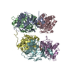
| |||||||||
|---|---|---|---|---|---|---|---|---|---|---|
| 1 | 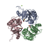
| |||||||||
| 2 | 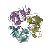
| |||||||||
| Unit cell |
| |||||||||
| Components on special symmetry positions |
| |||||||||
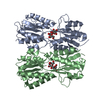
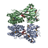
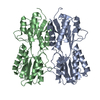
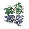
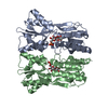
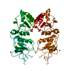
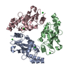
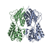
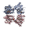
| Details | The biological assembly is a trimer, two of which are in the assymetric unit. Trimer 1 is made up of Chains A,B and C, Trimer 2 is made up of chains D, E and F. |
-Components
| #1: Protein | Mass: 17456.850 Da / Num. of mol.: 6 / Mutation: L80R Source method: isolated from a genetically manipulated source Source: (gene. exp.) Thermus thermophilus (bacteria) / Strain: HD8 / Plasmid: pET11a / Production host: References: UniProt: P83846, UniProt: Q5SIP7*PLUS, Transferases; Transferring one-carbon groups; Methyltransferases #2: Chemical | ChemComp-MG / |   Mass: 24.305 Da / Num. of mol.: 1 / Source method: obtained synthetically / Formula: Mg Mass: 24.305 Da / Num. of mol.: 1 / Source method: obtained synthetically / Formula: Mg#3: Chemical |   Mass: 35.453 Da / Num. of mol.: 2 / Source method: obtained synthetically / Formula: Cl Mass: 35.453 Da / Num. of mol.: 2 / Source method: obtained synthetically / Formula: Cl#4: Water | ChemComp-HOH / |  Mass: 18.015 Da / Num. of mol.: 398 / Source method: isolated from a natural source / Formula: H2O Mass: 18.015 Da / Num. of mol.: 398 / Source method: isolated from a natural source / Formula: H2OHas protein modification | Y | |
|---|
-Experimental details
-Experiment
| Experiment | Method: X-RAY DIFFRACTION / Number of used crystals: 1 |
|---|
- Sample preparation
| Crystal | Density Matthews: 2.12 Å3/Da / Density % sol: 41.46 % | |||||||||||||||||||||||||||||||||||||||||||||||||||||||||||||||
|---|---|---|---|---|---|---|---|---|---|---|---|---|---|---|---|---|---|---|---|---|---|---|---|---|---|---|---|---|---|---|---|---|---|---|---|---|---|---|---|---|---|---|---|---|---|---|---|---|---|---|---|---|---|---|---|---|---|---|---|---|---|---|---|---|
| Crystal grow | Temperature: 296 K / Method: vapor diffusion, sitting drop / pH: 8.3 Details: PEG 1000, magnesium chloride, Tris-Cl, pH 8.3, VAPOR DIFFUSION, SITTING DROP, temperature 296K | |||||||||||||||||||||||||||||||||||||||||||||||||||||||||||||||
| Crystal grow | *PLUS pH: 8 / Method: vapor diffusion, sitting drop | |||||||||||||||||||||||||||||||||||||||||||||||||||||||||||||||
| Components of the solutions | *PLUS
|
-Data collection
| Diffraction | Mean temperature: 100 K | ||||||||||||
|---|---|---|---|---|---|---|---|---|---|---|---|---|---|
| Diffraction source | Source: SYNCHROTRON / Site: SPring-8  / Beamline: BL26B1 / Wavelength: 0.97900, 0.97925, 0.97000 / Beamline: BL26B1 / Wavelength: 0.97900, 0.97925, 0.97000 | ||||||||||||
| Detector | Type: RIGAKU RAXIS V / Detector: IMAGE PLATE / Date: Jul 9, 2002 | ||||||||||||
| Radiation | Monochromator: Si 111 / Protocol: MAD / Monochromatic (M) / Laue (L): M / Scattering type: x-ray | ||||||||||||
| Radiation wavelength |
| ||||||||||||
| Reflection | Resolution: 2.3→38.5 Å / Num. all: 193179 / Num. obs: 191090 / % possible obs: 98.92 % / Observed criterion σ(F): 0 / Observed criterion σ(I): 0 / Redundancy: 5.05 % / Biso Wilson estimate: 22.4 Å2 / Rmerge(I) obs: 0.084 / Net I/σ(I): 15.96 | ||||||||||||
| Reflection shell | Resolution: 2.3→2.38 Å / Redundancy: 4.9 % / Rmerge(I) obs: 0.432 / Mean I/σ(I) obs: 3.32 / Num. unique all: 3762 / % possible all: 92.3 | ||||||||||||
| Reflection | *PLUS % possible obs: 90.8 % / Redundancy: 5.1 % | ||||||||||||
| Reflection shell | *PLUS % possible obs: 92.3 % / Redundancy: 4.9 % / Mean I/σ(I) obs: 3.3 |
- Processing
| Software |
| ||||||||||||||||||||||||||||||||||||
|---|---|---|---|---|---|---|---|---|---|---|---|---|---|---|---|---|---|---|---|---|---|---|---|---|---|---|---|---|---|---|---|---|---|---|---|---|---|
| Refinement | Method to determine structure: MAD / Resolution: 2.3→38.4 Å / Rfactor Rfree error: 0.004 / Data cutoff high absF: 231118.5 / Data cutoff low absF: 0 / Isotropic thermal model: RESTRAINED / Cross valid method: THROUGHOUT / σ(F): 0 / Stereochemistry target values: Engh & Huber Details: The following residues have been built into weak density and therefore should be interpretted with caution: A1, B160-164, C159-161, D158-163, E158-161 and F154:157
| ||||||||||||||||||||||||||||||||||||
| Solvent computation | Solvent model: FLAT MODEL / Bsol: 42.209 Å2 / ksol: 0.29942 e/Å3 | ||||||||||||||||||||||||||||||||||||
| Displacement parameters | Biso mean: 49.1 Å2
| ||||||||||||||||||||||||||||||||||||
| Refine analyze |
| ||||||||||||||||||||||||||||||||||||
| Refinement step | Cycle: LAST / Resolution: 2.3→38.4 Å
| ||||||||||||||||||||||||||||||||||||
| Refine LS restraints |
| ||||||||||||||||||||||||||||||||||||
| LS refinement shell | Resolution: 2.3→2.44 Å / Rfactor Rfree error: 0.012 / Total num. of bins used: 6
| ||||||||||||||||||||||||||||||||||||
| Xplor file |
| ||||||||||||||||||||||||||||||||||||
| Refinement | *PLUS Highest resolution: 2.3 Å / Lowest resolution: 38.5 Å / Rfactor Rfree: 0.279 / Rfactor Rwork: 0.217 | ||||||||||||||||||||||||||||||||||||
| Solvent computation | *PLUS | ||||||||||||||||||||||||||||||||||||
| Displacement parameters | *PLUS | ||||||||||||||||||||||||||||||||||||
| Refine LS restraints | *PLUS
| ||||||||||||||||||||||||||||||||||||
| LS refinement shell | *PLUS Rfactor Rfree: 0.323 |
