 Movie
Movie Controller


+ Open data

- Basic information
| Entry | Database: PDB / ID: 3lh4 | ||||||
|---|---|---|---|---|---|---|---|
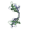
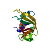
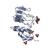
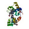
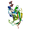
| Title | Crystal Structure of Sialostatin L2 | ||||||
 Components Components | Secreted cystatin | ||||||
 Keywords Keywords | HYDROLASE INHIBITOR / beta sheet / cystatin / Protease inhibitor / Thiol protease inhibitor | ||||||
| Function / homology |  Function and homology information Function and homology informationcysteine-type endopeptidase inhibitor activity / vesicle / : / cytoplasm Similarity search - Function | ||||||
| Biological species |  Ixodes scapularis (black-legged tick) Ixodes scapularis (black-legged tick) | ||||||
| Method |  X-RAY DIFFRACTION / SYNCHROTRON / MOLECULAR REPLACEMENT / Resolution: 1.8 Å X-RAY DIFFRACTION / SYNCHROTRON / MOLECULAR REPLACEMENT / Resolution: 1.8 Å | ||||||
 Authors Authors | Andersen, J.F. / Kotsyfakis, M. / Salat, J. / Horka, H. | ||||||
 Citation Citation | Journal: Mol.Microbiol. / Year: 2010 Title: The crystal structures of two salivary cystatins from the tick Ixodes scapularis and the effect of these inhibitors on the establishment of Borrelia burgdorferi infection in a murine model. Authors: Kotsyfakis, M. / Horka, H. / Salat, J. / Andersen, J.F. | ||||||
| History |
|
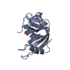
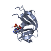
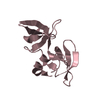
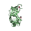
- Structure visualization
| Structure viewer | Molecule: MolmilJmol/JSmol |
|---|
- Downloads & links
-Download
| PDBx/mmCIF format | 3lh4.cif.gz | 38.1 KB | Display | PDBx/mmCIF format |
|---|---|---|---|---|
| PDB format | pdb3lh4.ent.gz | 26.2 KB | Display | PDB format |
| PDBx/mmJSON format | 3lh4.json.gz | Tree view | PDBx/mmJSON format | |
| Others |  Other downloads Other downloads |
-Validation report
| Arichive directory | https://data.pdbj.org/pub/pdb/validation_reports/lh/3lh4ftp://data.pdbj.org/pub/pdb/validation_reports/lh/3lh4 | HTTPS FTP |
|---|
-Related structure data


-Links
 PDBj
PDBj
- Assembly
| Deposited unit | 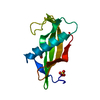
| ||||||||
|---|---|---|---|---|---|---|---|---|---|
| 1 |
| ||||||||
| Unit cell |
| ||||||||
| Components on special symmetry positions |
|
-Components
| #1: Protein | Mass: 12775.076 Da / Num. of mol.: 1 / Fragment: UNP residues 19-132 Source method: isolated from a genetically manipulated source Source: (gene. exp.) Ixodes scapularis (black-legged tick) / Gene: IscW_ISCW018602 / Plasmid: pET17b / Production host:  |
|---|---|
| #2: Chemical | ChemComp-SO4 /   Mass: 96.063 Da / Num. of mol.: 1 / Source method: obtained synthetically / Formula: SO4 Mass: 96.063 Da / Num. of mol.: 1 / Source method: obtained synthetically / Formula: SO4 |
| #3: Chemical | ChemComp-GOL /   Mass: 92.094 Da / Num. of mol.: 1 / Source method: obtained synthetically / Formula: C3H8O3 Mass: 92.094 Da / Num. of mol.: 1 / Source method: obtained synthetically / Formula: C3H8O3 |
| #4: Water | ChemComp-HOH /  Mass: 18.015 Da / Num. of mol.: 106 / Source method: isolated from a natural source / Formula: H2O Mass: 18.015 Da / Num. of mol.: 106 / Source method: isolated from a natural source / Formula: H2O |
| Has protein modification | Y |
-Experimental details
-Experiment
| Experiment | Method: X-RAY DIFFRACTION / Number of used crystals: 1 |
|---|
- Sample preparation
| Crystal | Density Matthews: 2.84 Å3/Da / Density % sol: 56.63 % |
|---|---|
| Crystal grow | Temperature: 298 K / Method: vapor diffusion, hanging drop / pH: 9 Details: 1.8 M Ammonium sulfate, 0.1 M Bicine, pH 9.0, VAPOR DIFFUSION, HANGING DROP, temperature 298K |
-Data collection
| Diffraction | Mean temperature: 100 K |
|---|---|
| Diffraction source | Source: SYNCHROTRON / Site: APS  / Beamline: 22-BM / Wavelength: 0.97902 Å / Beamline: 22-BM / Wavelength: 0.97902 Å |
| Detector | Type: MARMOSAIC 225 mm CCD / Detector: CCD / Date: Feb 21, 2008 |
| Radiation | Monochromator: Si(111) / Protocol: SINGLE WAVELENGTH / Monochromatic (M) / Laue (L): M / Scattering type: x-ray |
| Radiation wavelength | Wavelength: 0.97902 Å / Relative weight: 1 |
| Reflection | Resolution: 1.8→19.48 Å / Num. all: 13472 / Num. obs: 13472 / % possible obs: 99.8 % / Observed criterion σ(F): 0 / Observed criterion σ(I): 0 / Redundancy: 29.9 % / Rmerge(I) obs: 0.095 / Net I/σ(I): 22.1 |
| Reflection shell | Resolution: 1.8→1.86 Å / Redundancy: 28 % / Rmerge(I) obs: 0.195 / Mean I/σ(I) obs: 26.6 / Num. unique all: 1318 / % possible all: 100 |
- Processing
| Software |
| |||||||||||||||||||||||||||||||||||||||||||||||||||||||||||||||||
|---|---|---|---|---|---|---|---|---|---|---|---|---|---|---|---|---|---|---|---|---|---|---|---|---|---|---|---|---|---|---|---|---|---|---|---|---|---|---|---|---|---|---|---|---|---|---|---|---|---|---|---|---|---|---|---|---|---|---|---|---|---|---|---|---|---|---|
| Refinement | Method to determine structure: MOLECULAR REPLACEMENT / Resolution: 1.8→19.48 Å / Cor.coef. Fo:Fc: 0.958 / Cor.coef. Fo:Fc free: 0.954 / SU B: 4.357 / SU ML: 0.064 / Cross valid method: THROUGHOUT / σ(F): 0 / ESU R: 0.108 / ESU R Free: 0.102 / Stereochemistry target values: MAXIMUM LIKELIHOOD / Details: HYDROGENS HAVE BEEN ADDED IN THE RIDING POSITIONS
| |||||||||||||||||||||||||||||||||||||||||||||||||||||||||||||||||
| Solvent computation | Ion probe radii: 0.8 Å / Shrinkage radii: 0.8 Å / VDW probe radii: 1.4 Å / Solvent model: MASK | |||||||||||||||||||||||||||||||||||||||||||||||||||||||||||||||||
| Displacement parameters | Biso mean: 16.739 Å2 | |||||||||||||||||||||||||||||||||||||||||||||||||||||||||||||||||
| Refinement step | Cycle: LAST / Resolution: 1.8→19.48 Å
| |||||||||||||||||||||||||||||||||||||||||||||||||||||||||||||||||
| Refine LS restraints |
| |||||||||||||||||||||||||||||||||||||||||||||||||||||||||||||||||
| LS refinement shell | Resolution: 1.804→1.851 Å / Total num. of bins used: 20
| |||||||||||||||||||||||||||||||||||||||||||||||||||||||||||||||||
| Refinement TLS params. | Method: refined / Origin x: 32.2884 Å / Origin y: 24.2288 Å / Origin z: -1.2176 Å
|
