 Movie
Movie Controller

[English] 日本語
 Yorodumi
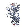
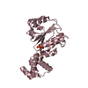
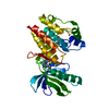
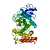
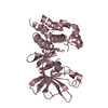
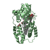
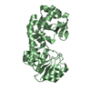
- PDB-7b8w: Structure of LIMK1 Kinase domain with allosteric inhibitor TH-470 -
+ Open data

- Basic information
| Entry | Database: PDB / ID: 7b8w | ||||||
|---|---|---|---|---|---|---|---|
| Title | Structure of LIMK1 Kinase domain with allosteric inhibitor TH-470 | ||||||
 Components Components | LIM domain kinase 1 | ||||||
 Keywords Keywords | SIGNALING PROTEIN / LIMK1 Kinase domain / inhibitor | ||||||
| Function / homology |  Function and homology information Function and homology informationpositive regulation of actin filament bundle assembly / RHO GTPases Activate ROCKs / axon extension / Sema4D induced cell migration and growth-cone collapse / stress fiber assembly / Fc-gamma receptor signaling pathway involved in phagocytosis / RHO GTPases activate PAKs / Sema3A PAK dependent Axon repulsion / ubiquitin ligase inhibitor activity / positive regulation of axon extension ...positive regulation of actin filament bundle assembly / RHO GTPases Activate ROCKs / axon extension / Sema4D induced cell migration and growth-cone collapse / stress fiber assembly / Fc-gamma receptor signaling pathway involved in phagocytosis / RHO GTPases activate PAKs / Sema3A PAK dependent Axon repulsion / ubiquitin ligase inhibitor activity / positive regulation of axon extension / Rho protein signal transduction / positive regulation of stress fiber assembly / heat shock protein binding / EPHB-mediated forward signaling / male germ cell nucleus / Regulation of actin dynamics for phagocytic cup formation / nervous system development / lamellipodium / actin cytoskeleton organization / cytoskeleton / protein phosphorylation / protein kinase activity / non-specific serine/threonine protein kinase / neuron projection / postsynapse / nuclear speck / protein serine kinase activity / focal adhesion / protein serine/threonine kinase activity / ubiquitin protein ligase binding / glutamatergic synapse / signal transduction / ATP binding / metal ion binding / membrane / nucleus / cytoplasm / cytosol Similarity search - Function | ||||||
| Biological species |  Homo sapiens (human) Homo sapiens (human) | ||||||
| Method |  X-RAY DIFFRACTION / SYNCHROTRON / MOLECULAR REPLACEMENT / molecular replacement / Resolution: 2.8 Å X-RAY DIFFRACTION / SYNCHROTRON / MOLECULAR REPLACEMENT / molecular replacement / Resolution: 2.8 Å | ||||||
 Authors Authors | Lee, H. / Yosaatmadja, Y. / Burgess-Brown, N.A. / von Delft, F. / Arrowsmith, C.H. / Edwards, A. / Bountra, C. / Elkins, J.M. | ||||||
| Funding support |  United Kingdom, 1items United Kingdom, 1items
| ||||||
 Citation Citation | Journal: To Be Published Title: Structure of LIMK1 Kinase domain with allosteric inhibitor TH-470 Authors: Lee, H. / Yosaatmadja, Y. / Burgess-Brown, N.A. / von Delft, F. / Arrowsmith, C.H. / Edwards, A. / Bountra, C. / Elkins, J.M. | ||||||
| History |
|
- Structure visualization
| Structure viewer | Molecule: MolmilJmol/JSmol |
|---|
- Downloads & links
-Download
| PDBx/mmCIF format | 7b8w.cif.gz | 226.7 KB | Display | PDBx/mmCIF format |
|---|---|---|---|---|
| PDB format | pdb7b8w.ent.gz | 179.1 KB | Display | PDB format |
| PDBx/mmJSON format | 7b8w.json.gz | Tree view | PDBx/mmJSON format | |
| Others |  Other downloads Other downloads |
-Validation report
| Arichive directory | https://data.pdbj.org/pub/pdb/validation_reports/b8/7b8wftp://data.pdbj.org/pub/pdb/validation_reports/b8/7b8w | HTTPS FTP |
|---|
-Related structure data
| Related structure data | 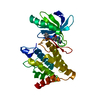 5nxcS S: Starting model for refinement |
|---|---|
| Similar structure data |
-Links
 PDBj
PDBj


- Assembly
| Deposited unit | 
| |||||||||||||||||||||||||||||||||||||||||||||||||||||||||||||||||
|---|---|---|---|---|---|---|---|---|---|---|---|---|---|---|---|---|---|---|---|---|---|---|---|---|---|---|---|---|---|---|---|---|---|---|---|---|---|---|---|---|---|---|---|---|---|---|---|---|---|---|---|---|---|---|---|---|---|---|---|---|---|---|---|---|---|---|
| 1 | 
| |||||||||||||||||||||||||||||||||||||||||||||||||||||||||||||||||
| 2 | 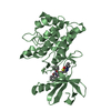
| |||||||||||||||||||||||||||||||||||||||||||||||||||||||||||||||||
| 3 | 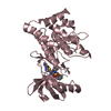
| |||||||||||||||||||||||||||||||||||||||||||||||||||||||||||||||||
| 4 | 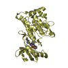
| |||||||||||||||||||||||||||||||||||||||||||||||||||||||||||||||||
| Unit cell |
| |||||||||||||||||||||||||||||||||||||||||||||||||||||||||||||||||
| Noncrystallographic symmetry (NCS) | NCS domain:
NCS domain segments:
NCS oper:
|
-Components
| #1: Protein | Mass: 35994.773 Da / Num. of mol.: 4 Source method: isolated from a genetically manipulated source Source: (gene. exp.) Homo sapiens (human) / Gene: LIMK1, LIMK / Production host:   Spodoptera frugiperda (fall armyworm) Spodoptera frugiperda (fall armyworm)References: UniProt: P53667, non-specific serine/threonine protein kinase #2: Chemical | ChemComp-T3B /   Mass: 605.728 Da / Num. of mol.: 4 / Source method: obtained synthetically / Formula: C30H31N5O5S2 / Feature type: SUBJECT OF INVESTIGATION Mass: 605.728 Da / Num. of mol.: 4 / Source method: obtained synthetically / Formula: C30H31N5O5S2 / Feature type: SUBJECT OF INVESTIGATION#3: Chemical |   Mass: 62.068 Da / Num. of mol.: 3 / Source method: obtained synthetically / Formula: C2H6O2 Mass: 62.068 Da / Num. of mol.: 3 / Source method: obtained synthetically / Formula: C2H6O2#4: Water | ChemComp-HOH / |  Mass: 18.015 Da / Num. of mol.: 8 / Source method: isolated from a natural source / Formula: H2O Mass: 18.015 Da / Num. of mol.: 8 / Source method: isolated from a natural source / Formula: H2OHas ligand of interest | Y | |
|---|
-Experimental details
-Experiment
| Experiment | Method: X-RAY DIFFRACTION / Number of used crystals: 1 |
|---|
- Sample preparation
| Crystal | Density Matthews: 2.38 Å3/Da / Density % sol: 48.33 % |
|---|---|
| Crystal grow | Temperature: 277.15 K / Method: vapor diffusion, sitting drop / Details: 0.2M Ammonium Citrate dibasic, 20% w/v PEG 3350 |
-Data collection
| Diffraction | Mean temperature: 100 K / Serial crystal experiment: N | ||||||||||||||||||||||||||||||
|---|---|---|---|---|---|---|---|---|---|---|---|---|---|---|---|---|---|---|---|---|---|---|---|---|---|---|---|---|---|---|---|
| Diffraction source | Source: SYNCHROTRON / Site: Diamond / Beamline: I24 / Wavelength: 0.9686 Å | ||||||||||||||||||||||||||||||
| Detector | Type: DECTRIS PILATUS3 6M / Detector: PIXEL / Date: Feb 28, 2019 | ||||||||||||||||||||||||||||||
| Radiation | Protocol: SINGLE WAVELENGTH / Monochromatic (M) / Laue (L): M / Scattering type: x-ray | ||||||||||||||||||||||||||||||
| Radiation wavelength | Wavelength: 0.9686 Å / Relative weight: 1 | ||||||||||||||||||||||||||||||
| Reflection | Resolution: 2.8→51.2 Å / Num. obs: 33406 / % possible obs: 99.9 % / Redundancy: 6.8 % / CC1/2: 0.987 / Rmerge(I) obs: 0.221 / Rpim(I) all: 0.091 / Rrim(I) all: 0.239 / Net I/σ(I): 7 / Num. measured all: 225877 / Scaling rejects: 24 | ||||||||||||||||||||||||||||||
| Reflection shell | Diffraction-ID: 1
|
-Phasing
| Phasing | Method: molecular replacement | |||||||||
|---|---|---|---|---|---|---|---|---|---|---|
| Phasing MR | Model details: Phaser MODE: MR_AUTO
|
- Processing
| Software |
| ||||||||||||||||||||||||||||||||||||||||||||||||||||||||||||
|---|---|---|---|---|---|---|---|---|---|---|---|---|---|---|---|---|---|---|---|---|---|---|---|---|---|---|---|---|---|---|---|---|---|---|---|---|---|---|---|---|---|---|---|---|---|---|---|---|---|---|---|---|---|---|---|---|---|---|---|---|---|
| Refinement | Method to determine structure: MOLECULAR REPLACEMENT Starting model: 5NXC Resolution: 2.8→50.15 Å / Cor.coef. Fo:Fc: 0.932 / Cor.coef. Fo:Fc free: 0.858 / SU B: 18.604 / SU ML: 0.352 / Cross valid method: THROUGHOUT / σ(F): 0 / ESU R Free: 0.425 / Stereochemistry target values: MAXIMUM LIKELIHOOD Details: HYDROGENS HAVE BEEN ADDED IN THE RIDING POSITIONS U VALUES : REFINED INDIVIDUALLY
| ||||||||||||||||||||||||||||||||||||||||||||||||||||||||||||
| Solvent computation | Ion probe radii: 0.8 Å / Shrinkage radii: 0.8 Å / VDW probe radii: 1.2 Å / Solvent model: MASK | ||||||||||||||||||||||||||||||||||||||||||||||||||||||||||||
| Displacement parameters | Biso max: 118.53 Å2 / Biso mean: 45.413 Å2 / Biso min: 24.34 Å2
| ||||||||||||||||||||||||||||||||||||||||||||||||||||||||||||
| Refinement step | Cycle: final / Resolution: 2.8→50.15 Å
| ||||||||||||||||||||||||||||||||||||||||||||||||||||||||||||
| Refine LS restraints |
| ||||||||||||||||||||||||||||||||||||||||||||||||||||||||||||
| Refine LS restraints NCS | Ens-ID: 1 / Number: 3903 / Refine-ID: X-RAY DIFFRACTION
| ||||||||||||||||||||||||||||||||||||||||||||||||||||||||||||
| LS refinement shell | Resolution: 2.8→2.873 Å / Rfactor Rfree error: 0 / Total num. of bins used: 20
|
