 Movie
Movie Controller

[English] 日本語
 Yorodumi
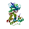
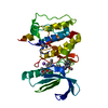
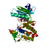
- PDB-2fum: Catalytic domain of protein kinase PknB from Mycobacterium tuberc... -
+ Open data

- Basic information
| Entry | Database: PDB / ID: 2fum | ||||||
|---|---|---|---|---|---|---|---|
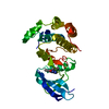
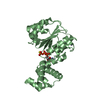
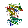
| Title | Catalytic domain of protein kinase PknB from Mycobacterium tuberculosis in complex with mitoxantrone | ||||||
 Components Components | Probable serine/threonine-protein kinase pknB | ||||||
 Keywords Keywords | TRANSFERASE / PROTEIN KINASE-INHIBITOR COMPLEX | ||||||
| Function / homology |  Function and homology information Function and homology informationnegative regulation of growth rate / negative regulation of fatty acid biosynthetic process / membrane => GO:0016020 / peptidoglycan biosynthetic process / protein serine/threonine/tyrosine kinase activity / peptidoglycan-based cell wall / regulation of cell shape / manganese ion binding / protein kinase activity / non-specific serine/threonine protein kinase ...negative regulation of growth rate / negative regulation of fatty acid biosynthetic process / membrane => GO:0016020 / peptidoglycan biosynthetic process / protein serine/threonine/tyrosine kinase activity / peptidoglycan-based cell wall / regulation of cell shape / manganese ion binding / protein kinase activity / non-specific serine/threonine protein kinase / protein serine kinase activity / protein serine/threonine kinase activity / regulation of DNA-templated transcription / ATP binding / metal ion binding / identical protein binding / plasma membrane Similarity search - Function | ||||||
| Biological species |  Mycobacterium tuberculosis H37Rv (bacteria) Mycobacterium tuberculosis H37Rv (bacteria) | ||||||
| Method |  X-RAY DIFFRACTION / SYNCHROTRON / MOLECULAR REPLACEMENT / Resolution: 2.89 Å X-RAY DIFFRACTION / SYNCHROTRON / MOLECULAR REPLACEMENT / Resolution: 2.89 Å | ||||||
 Authors Authors | Wehenkel, A. / Alzari, P.M. | ||||||
 Citation Citation | Journal: Febs Lett. / Year: 2006 Title: The structure of PknB in complex with mitoxantrone, an ATP-competitive inhibitor, suggests a mode of protein kinase regulation in mycobacteria Authors: Wehenkel, A. / Fernandez, P. / Bellinzoni, M. / Catherinot, V. / Barilone, N. / Labesse, G. / Jackson, M. / Alzari, P.M. | ||||||
| History |
|
- Structure visualization
| Structure viewer | Molecule: MolmilJmol/JSmol |
|---|
- Downloads & links
-Download
| PDBx/mmCIF format | 2fum.cif.gz | 204.6 KB | Display | PDBx/mmCIF format |
|---|---|---|---|---|
| PDB format | pdb2fum.ent.gz | 164 KB | Display | PDB format |
| PDBx/mmJSON format | 2fum.json.gz | Tree view | PDBx/mmJSON format | |
| Others |  Other downloads Other downloads |
-Validation report
| Arichive directory | https://data.pdbj.org/pub/pdb/validation_reports/fu/2fumftp://data.pdbj.org/pub/pdb/validation_reports/fu/2fum | HTTPS FTP |
|---|
-Related structure data
| Related structure data | 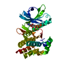 1o6yS S: Starting model for refinement |
|---|---|
| Similar structure data |
-Links
 PDBj
PDBj- Assembly
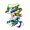
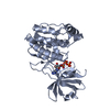
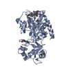
| Deposited unit | 
| |||||||||||||||||||||||||||||||||||||||||||||||||||||||||||||||||||||||||||||||||||||||||||||||||||||||||||||||||||||||||||||||||
|---|---|---|---|---|---|---|---|---|---|---|---|---|---|---|---|---|---|---|---|---|---|---|---|---|---|---|---|---|---|---|---|---|---|---|---|---|---|---|---|---|---|---|---|---|---|---|---|---|---|---|---|---|---|---|---|---|---|---|---|---|---|---|---|---|---|---|---|---|---|---|---|---|---|---|---|---|---|---|---|---|---|---|---|---|---|---|---|---|---|---|---|---|---|---|---|---|---|---|---|---|---|---|---|---|---|---|---|---|---|---|---|---|---|---|---|---|---|---|---|---|---|---|---|---|---|---|---|---|---|---|
| 1 | 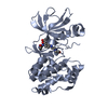
| |||||||||||||||||||||||||||||||||||||||||||||||||||||||||||||||||||||||||||||||||||||||||||||||||||||||||||||||||||||||||||||||||
| 2 | 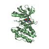
| |||||||||||||||||||||||||||||||||||||||||||||||||||||||||||||||||||||||||||||||||||||||||||||||||||||||||||||||||||||||||||||||||
| 3 | 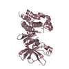
| |||||||||||||||||||||||||||||||||||||||||||||||||||||||||||||||||||||||||||||||||||||||||||||||||||||||||||||||||||||||||||||||||
| 4 | 
| |||||||||||||||||||||||||||||||||||||||||||||||||||||||||||||||||||||||||||||||||||||||||||||||||||||||||||||||||||||||||||||||||
| Unit cell |
| |||||||||||||||||||||||||||||||||||||||||||||||||||||||||||||||||||||||||||||||||||||||||||||||||||||||||||||||||||||||||||||||||
| Noncrystallographic symmetry (NCS) | NCS domain:
NCS domain segments: Refine code: 2
NCS ensembles :
|
-Components
| #1: Protein | Mass: 32458.506 Da / Num. of mol.: 4 / Fragment: CATALYTIC DOMAIN Source method: isolated from a genetically manipulated source Source: (gene. exp.) Mycobacterium tuberculosis H37Rv (bacteria)Species: Mycobacterium tuberculosis / Strain: H37RV / Gene: pknB / Plasmid: PET28 / Species (production host): Escherichia coli / Production host: References: UniProt: P0A5S4, UniProt: P9WI81*PLUS, EC: 2.7.1.37 #2: Chemical | ChemComp-MIX / 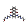  Mass: 444.481 Da / Num. of mol.: 4 / Source method: obtained synthetically / Formula: C22H28N4O6 / Comment: antineoplastic*YM Mass: 444.481 Da / Num. of mol.: 4 / Source method: obtained synthetically / Formula: C22H28N4O6 / Comment: antineoplastic*YM |
|---|
-Experimental details
-Experiment
| Experiment | Method: X-RAY DIFFRACTION / Number of used crystals: 1 |
|---|
- Sample preparation
| Crystal | Density Matthews: 3.42 Å3/Da / Density % sol: 64.05 % |
|---|---|
| Crystal grow | Temperature: 291 K / Method: vapor diffusion, hanging drop / pH: 5.6 Details: 1.2M NaAcetate 50mM NaCacodylate, pH 5.6, VAPOR DIFFUSION, HANGING DROP, temperature 291K |
-Data collection
| Diffraction | Mean temperature: 100 K |
|---|---|
| Diffraction source | Source: SYNCHROTRON / Site: ESRF  / Beamline: ID14-3 / Wavelength: 0.931 Å / Beamline: ID14-3 / Wavelength: 0.931 Å |
| Detector | Type: MARRESEARCH / Detector: CCD / Date: Mar 10, 2005 |
| Radiation | Monochromator: Diamond (111), Ge(220) / Protocol: SINGLE WAVELENGTH / Monochromatic (M) / Laue (L): M / Scattering type: x-ray |
| Radiation wavelength | Wavelength: 0.931 Å / Relative weight: 1 |
| Reflection | Resolution: 2.89→69.84 Å / Num. all: 28195 / Num. obs: 28195 / % possible obs: 63.47 % / Observed criterion σ(F): 1 / Observed criterion σ(I): 1 |
| Reflection shell | Resolution: 2.89→3.21 Å / % possible all: 16 |
- Processing
| Software |
| |||||||||||||||||||||||||||||||||||||||||||||||||||||||||||||||||||||||||||||||||||||||||||||||||||||||||||||||||||||||||||||||||||||||||||||||||||||||||||||||||||||||||||||||||||||||||||||||||||||||||||||||||||||||||||||||||
|---|---|---|---|---|---|---|---|---|---|---|---|---|---|---|---|---|---|---|---|---|---|---|---|---|---|---|---|---|---|---|---|---|---|---|---|---|---|---|---|---|---|---|---|---|---|---|---|---|---|---|---|---|---|---|---|---|---|---|---|---|---|---|---|---|---|---|---|---|---|---|---|---|---|---|---|---|---|---|---|---|---|---|---|---|---|---|---|---|---|---|---|---|---|---|---|---|---|---|---|---|---|---|---|---|---|---|---|---|---|---|---|---|---|---|---|---|---|---|---|---|---|---|---|---|---|---|---|---|---|---|---|---|---|---|---|---|---|---|---|---|---|---|---|---|---|---|---|---|---|---|---|---|---|---|---|---|---|---|---|---|---|---|---|---|---|---|---|---|---|---|---|---|---|---|---|---|---|---|---|---|---|---|---|---|---|---|---|---|---|---|---|---|---|---|---|---|---|---|---|---|---|---|---|---|---|---|---|---|---|---|---|---|---|---|---|---|---|---|---|---|---|---|---|---|---|---|
| Refinement | Method to determine structure: MOLECULAR REPLACEMENT Starting model: PDB ENTRY 1O6Y Resolution: 2.89→69.84 Å / Cor.coef. Fo:Fc: 0.938 / Cor.coef. Fo:Fc free: 0.905 / SU B: 53.161 / SU ML: 0.412 / TLS residual ADP flag: LIKELY RESIDUAL / Cross valid method: THROUGHOUT / σ(F): 0 / ESU R Free: 0.509 / Stereochemistry target values: MAXIMUM LIKELIHOOD Details: THE DATA IS HIGHLY ANISOTROPIC (2.89 A RESOLUTION LIMIT ALONG THE c* AXIS, BUT ONLY 3.5 A ALONG THE a* AND b* AXES), ACCOUNTING FOR THE POOR DATA COMPLETENESS AT THE HIGHER RESOLUTION SHELLS ...Details: THE DATA IS HIGHLY ANISOTROPIC (2.89 A RESOLUTION LIMIT ALONG THE c* AXIS, BUT ONLY 3.5 A ALONG THE a* AND b* AXES), ACCOUNTING FOR THE POOR DATA COMPLETENESS AT THE HIGHER RESOLUTION SHELLS (DATA IS 99% COMPLETE AT 3.49 A, 45% IN THE 3.49-3.21 A SHELL, AND ONLY 16% IN THE 3.21-2.89 A SHELL.
| |||||||||||||||||||||||||||||||||||||||||||||||||||||||||||||||||||||||||||||||||||||||||||||||||||||||||||||||||||||||||||||||||||||||||||||||||||||||||||||||||||||||||||||||||||||||||||||||||||||||||||||||||||||||||||||||||
| Solvent computation | Ion probe radii: 0.8 Å / Shrinkage radii: 0.8 Å / VDW probe radii: 1.2 Å / Solvent model: MASK | |||||||||||||||||||||||||||||||||||||||||||||||||||||||||||||||||||||||||||||||||||||||||||||||||||||||||||||||||||||||||||||||||||||||||||||||||||||||||||||||||||||||||||||||||||||||||||||||||||||||||||||||||||||||||||||||||
| Displacement parameters | Biso mean: 79.784 Å2
| |||||||||||||||||||||||||||||||||||||||||||||||||||||||||||||||||||||||||||||||||||||||||||||||||||||||||||||||||||||||||||||||||||||||||||||||||||||||||||||||||||||||||||||||||||||||||||||||||||||||||||||||||||||||||||||||||
| Refinement step | Cycle: LAST / Resolution: 2.89→69.84 Å
| |||||||||||||||||||||||||||||||||||||||||||||||||||||||||||||||||||||||||||||||||||||||||||||||||||||||||||||||||||||||||||||||||||||||||||||||||||||||||||||||||||||||||||||||||||||||||||||||||||||||||||||||||||||||||||||||||
| Refine LS restraints |
| |||||||||||||||||||||||||||||||||||||||||||||||||||||||||||||||||||||||||||||||||||||||||||||||||||||||||||||||||||||||||||||||||||||||||||||||||||||||||||||||||||||||||||||||||||||||||||||||||||||||||||||||||||||||||||||||||
| Refine LS restraints NCS | Dom-ID: 1 / Refine-ID: X-RAY DIFFRACTION
| |||||||||||||||||||||||||||||||||||||||||||||||||||||||||||||||||||||||||||||||||||||||||||||||||||||||||||||||||||||||||||||||||||||||||||||||||||||||||||||||||||||||||||||||||||||||||||||||||||||||||||||||||||||||||||||||||
| LS refinement shell | Resolution: 2.891→2.966 Å / Total num. of bins used: 20
| |||||||||||||||||||||||||||||||||||||||||||||||||||||||||||||||||||||||||||||||||||||||||||||||||||||||||||||||||||||||||||||||||||||||||||||||||||||||||||||||||||||||||||||||||||||||||||||||||||||||||||||||||||||||||||||||||
| Refinement TLS params. | Method: refined / Refine-ID: X-RAY DIFFRACTION
| |||||||||||||||||||||||||||||||||||||||||||||||||||||||||||||||||||||||||||||||||||||||||||||||||||||||||||||||||||||||||||||||||||||||||||||||||||||||||||||||||||||||||||||||||||||||||||||||||||||||||||||||||||||||||||||||||
| Refinement TLS group |
|
