 Movie
Movie Controller

[English] 日本語
 Yorodumi
- PDB-4fg8: Crystal structure of human calcium/calmodulin-dependent protein k... -
+ Open data

- Basic information
| Entry | Database: PDB / ID: 4fg8 | ||||||
|---|---|---|---|---|---|---|---|
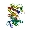
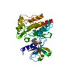
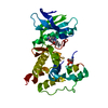
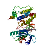
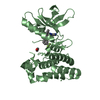
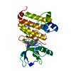
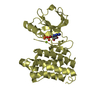
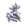
| Title | Crystal structure of human calcium/calmodulin-dependent protein kinase I 1-315 in complex with ATP | ||||||
 Components Components | Calcium/calmodulin-dependent protein kinase type 1 | ||||||
 Keywords Keywords | TRANSFERASE / CaMK / calmodulin / autoinhibition / regulation mechanism / kinase | ||||||
| Function / homology |  Function and homology information Function and homology informationpositive regulation of syncytium formation by plasma membrane fusion / positive regulation of synapse structural plasticity / regulation of muscle cell differentiation / Ca2+/calmodulin-dependent protein kinase / calcium/calmodulin-dependent protein kinase activity / nucleocytoplasmic transport / positive regulation of muscle cell differentiation / Activation of RAC1 downstream of NMDARs / regulation of synapse organization / positive regulation of dendritic spine development ...positive regulation of syncytium formation by plasma membrane fusion / positive regulation of synapse structural plasticity / regulation of muscle cell differentiation / Ca2+/calmodulin-dependent protein kinase / calcium/calmodulin-dependent protein kinase activity / nucleocytoplasmic transport / positive regulation of muscle cell differentiation / Activation of RAC1 downstream of NMDARs / regulation of synapse organization / positive regulation of dendritic spine development / Negative regulation of NMDA receptor-mediated neuronal transmission / positive regulation of protein export from nucleus / positive regulation of neuron projection development / nervous system development / regulation of protein localization / cell differentiation / calmodulin binding / postsynaptic density / intracellular signal transduction / protein serine kinase activity / protein serine/threonine kinase activity / signal transduction / positive regulation of transcription by RNA polymerase II / ATP binding / nucleus / cytoplasm / cytosol Similarity search - Function | ||||||
| Biological species |  Homo sapiens (human) Homo sapiens (human) | ||||||
| Method |  X-RAY DIFFRACTION / SYNCHROTRON / MOLECULAR REPLACEMENT / Resolution: 2.2 Å X-RAY DIFFRACTION / SYNCHROTRON / MOLECULAR REPLACEMENT / Resolution: 2.2 Å | ||||||
 Authors Authors | Zha, M. / Zhong, C. / Ou, Y. / Wang, J. / Han, L. / Ding, J. | ||||||
 Citation Citation | Journal: Plos One / Year: 2012 Title: Crystal structures of human CaMKIalpha reveal insights into the regulation mechanism of CaMKI. Authors: Zha, M. / Zhong, C. / Ou, Y. / Han, L. / Wang, J. / Ding, J. | ||||||
| History |
|
- Structure visualization
| Structure viewer | Molecule: MolmilJmol/JSmol |
|---|
- Downloads & links
-Download
| PDBx/mmCIF format | 4fg8.cif.gz | 225 KB | Display | PDBx/mmCIF format |
|---|---|---|---|---|
| PDB format | pdb4fg8.ent.gz | 179.9 KB | Display | PDB format |
| PDBx/mmJSON format | 4fg8.json.gz | Tree view | PDBx/mmJSON format | |
| Others |  Other downloads Other downloads |
-Validation report
| Arichive directory | https://data.pdbj.org/pub/pdb/validation_reports/fg/4fg8ftp://data.pdbj.org/pub/pdb/validation_reports/fg/4fg8 | HTTPS FTP |
|---|
-Related structure data
| Related structure data |  4fg7SC  4fg9C  4fgbC S: Starting model for refinement C: citing same article ( |
|---|---|
| Similar structure data |


-Links
 PDBj
PDBj


- Assembly
| Deposited unit | 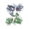
| ||||||||
|---|---|---|---|---|---|---|---|---|---|
| 1 | 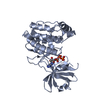
| ||||||||
| 2 | 
| ||||||||
| Unit cell |
|
-Components
| #1: Protein | Mass: 35765.699 Da / Num. of mol.: 2 Source method: isolated from a genetically manipulated source Source: (gene. exp.) Homo sapiens (human) / Gene: CAMK1 / Plasmid: pGEX4T-1 / Production host:  References: UniProt: Q14012, Ca2+/calmodulin-dependent protein kinase #2: Chemical |   Mass: 507.181 Da / Num. of mol.: 2 / Source method: obtained synthetically / Formula: C10H16N5O13P3 / Comment: ATP, energy-carrying molecule*YM Mass: 507.181 Da / Num. of mol.: 2 / Source method: obtained synthetically / Formula: C10H16N5O13P3 / Comment: ATP, energy-carrying molecule*YM#3: Water | ChemComp-HOH / |  Mass: 18.015 Da / Num. of mol.: 135 / Source method: isolated from a natural source / Formula: H2O Mass: 18.015 Da / Num. of mol.: 135 / Source method: isolated from a natural source / Formula: H2O |
|---|
-Experimental details
-Experiment
| Experiment | Method: X-RAY DIFFRACTION / Number of used crystals: 1 |
|---|
- Sample preparation
| Crystal | Density Matthews: 2.15 Å3/Da / Density % sol: 42.88 % |
|---|---|
| Crystal grow | Temperature: 277 K / Method: vapor diffusion, hanging drop / pH: 6 Details: 3% dioxane, 100mM MES, 1.5M (NH4)2SO4, pH 6.0, vapor diffusion, hanging drop, temperature 277K |
-Data collection
| Diffraction | Mean temperature: 100 K | |||||||||||||||
|---|---|---|---|---|---|---|---|---|---|---|---|---|---|---|---|---|
| Diffraction source | Source: SYNCHROTRON / Site: SSRF  / Beamline: BL17U / Wavelength: 0.9791 Å / Beamline: BL17U / Wavelength: 0.9791 Å | |||||||||||||||
| Detector | Type: RAYONIX MX315HE / Detector: CCD / Date: Jan 13, 2012 | |||||||||||||||
| Radiation | Protocol: SINGLE WAVELENGTH / Monochromatic (M) / Laue (L): M / Scattering type: x-ray | |||||||||||||||
| Radiation wavelength | Wavelength: 0.9791 Å / Relative weight: 1 | |||||||||||||||
| Reflection twin |
| |||||||||||||||
| Reflection | Resolution: 2.2→76.7 Å / Num. all: 30376 / % possible obs: 99.1 % / Redundancy: 5.5 % / Biso Wilson estimate: 43.4 Å2 / Rmerge(I) obs: 0.072 / Net I/σ(I): 22.4 | |||||||||||||||
| Reflection shell | Resolution: 2.2→2.28 Å / % possible all: 99.4 |
- Processing
| Software |
| ||||||||||||||||||||||||||||||||||||||||||||||||||||||||||||||||||||||
|---|---|---|---|---|---|---|---|---|---|---|---|---|---|---|---|---|---|---|---|---|---|---|---|---|---|---|---|---|---|---|---|---|---|---|---|---|---|---|---|---|---|---|---|---|---|---|---|---|---|---|---|---|---|---|---|---|---|---|---|---|---|---|---|---|---|---|---|---|---|---|---|
| Refinement | Method to determine structure: MOLECULAR REPLACEMENT Starting model: PDB ENTRY 4FG7 Resolution: 2.2→76.7 Å / Cor.coef. Fo:Fc: 0.952 / Cor.coef. Fo:Fc free: 0.921 / Occupancy max: 1 / Occupancy min: 1 / SU ML: 0.15 / Cross valid method: THROUGHOUT / σ(F): 0 / ESU R Free: 0.046 / Stereochemistry target values: MAXIMUM LIKELIHOOD / Details: U VALUES : REFINED INDIVIDUALLY
| ||||||||||||||||||||||||||||||||||||||||||||||||||||||||||||||||||||||
| Solvent computation | Ion probe radii: 0.8 Å / Shrinkage radii: 0.8 Å / VDW probe radii: 1.4 Å / Solvent model: MASK | ||||||||||||||||||||||||||||||||||||||||||||||||||||||||||||||||||||||
| Displacement parameters | Biso max: 184.56 Å2 / Biso mean: 54.5095 Å2 / Biso min: 30.41 Å2 | ||||||||||||||||||||||||||||||||||||||||||||||||||||||||||||||||||||||
| Refinement step | Cycle: LAST / Resolution: 2.2→76.7 Å
| ||||||||||||||||||||||||||||||||||||||||||||||||||||||||||||||||||||||
| Refine LS restraints |
| ||||||||||||||||||||||||||||||||||||||||||||||||||||||||||||||||||||||
| LS refinement shell | Resolution: 2.199→2.256 Å / Total num. of bins used: 20
|
