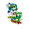
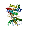
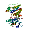
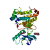
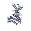
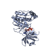
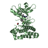
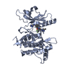
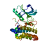
| Entry | Database: PDB / ID: 4aw5
|
|---|
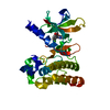
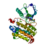
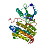
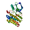
| Title | Complex of the EphB4 kinase domain with an oxindole inhibitor |
|---|
 Components Components | EPHRIN TYPE-B RECEPTOR 4 |
|---|
 Keywords Keywords | TRANSFERASE |
|---|
| Function / homology |  Function and homology information Function and homology information
ephrin receptor activity / cell migration involved in sprouting angiogenesis / EPH-Ephrin signaling / Ephrin signaling / EPH-ephrin mediated repulsion of cells / ephrin receptor signaling pathway / heart morphogenesis / EPHB-mediated forward signaling / transmembrane receptor protein tyrosine kinase activity / receptor protein-tyrosine kinase ...ephrin receptor activity / cell migration involved in sprouting angiogenesis / EPH-Ephrin signaling / Ephrin signaling / EPH-ephrin mediated repulsion of cells / ephrin receptor signaling pathway / heart morphogenesis / EPHB-mediated forward signaling / transmembrane receptor protein tyrosine kinase activity / receptor protein-tyrosine kinase / angiogenesis / cell adhesion / signaling receptor complex / extracellular exosome / extracellular region / ATP binding / plasma membrane / cytosolSimilarity search - Function Ephrin type-B receptor 4, ligand binding domain / EPH-B4, SAM domain / Tyrosine-protein kinase ephrin type A/B receptor-like / Tyrosine-protein kinase ephrin type A/B receptor-like / Ephrin cysteine rich domain / Ephrin receptor type-A /type-B / Ephrin receptor ligand binding domain / Tyrosine-protein kinase, receptor class V, conserved site / Ephrin receptor, transmembrane domain / : ...Ephrin type-B receptor 4, ligand binding domain / EPH-B4, SAM domain / Tyrosine-protein kinase ephrin type A/B receptor-like / Tyrosine-protein kinase ephrin type A/B receptor-like / Ephrin cysteine rich domain / Ephrin receptor type-A /type-B / Ephrin receptor ligand binding domain / Tyrosine-protein kinase, receptor class V, conserved site / Ephrin receptor, transmembrane domain / : / Ephrin receptor ligand binding domain / Ephrin type-A receptor 2 transmembrane domain / Receptor tyrosine kinase class V signature 1. / Receptor tyrosine kinase class V signature 2. / Eph receptor ligand-binding domain profile. / Ephrin receptor ligand binding domain / Putative ephrin-receptor like / SAM domain (Sterile alpha motif) / SAM domain profile. / Sterile alpha motif. / Sterile alpha motif domain / Sterile alpha motif/pointed domain superfamily / Fibronectin type III domain / Growth factor receptor cysteine-rich domain superfamily / EGF-like domain signature 2. / Fibronectin type 3 domain / Fibronectin type-III domain profile. / Galactose-binding-like domain superfamily / Fibronectin type III / Fibronectin type III superfamily / Tyrosine-protein kinase, catalytic domain / Tyrosine kinase, catalytic domain / Tyrosine protein kinases specific active-site signature. / Tyrosine-protein kinase, active site / Protein tyrosine and serine/threonine kinase / Serine-threonine/tyrosine-protein kinase, catalytic domain / Phosphorylase Kinase; domain 1 / Phosphorylase Kinase; domain 1 / Transferase(Phosphotransferase) domain 1 / Transferase(Phosphotransferase); domain 1 / Protein kinase, ATP binding site / Protein kinases ATP-binding region signature. / Immunoglobulin-like fold / Protein kinase domain profile. / Protein kinase domain / Protein kinase-like domain superfamily / 2-Layer Sandwich / Orthogonal Bundle / Mainly Alpha / Alpha BetaSimilarity search - Domain/homology |
|---|
| Biological species |  HOMO SAPIENS (human) HOMO SAPIENS (human) |
|---|
| Method |  X-RAY DIFFRACTION / SYNCHROTRON / MOLECULAR REPLACEMENT / Resolution: 2.33 Å X-RAY DIFFRACTION / SYNCHROTRON / MOLECULAR REPLACEMENT / Resolution: 2.33 Å |
|---|
 Authors Authors | Till, J.H. / Stout, T.J. |
|---|
 Citation Citation | Journal: Bioorg.Med.Chem.Lett. / Year: 2012
Title: The Design, Synthesis, and Biological Evaluation of Potent Receptor Tyrosine Kinase Inhibitors.
Authors: Kim, M.H. / Tsuhako, A.L. / Co, E.W. / Aftab, D.T. / Bentzien, F. / Chen, J. / Cheng, W. / Engst, S. / Goon, L. / Klein, R.R. / Le, D.T. / Mac, M. / Parks, J.J. / Qian, F. / Rodriquez, M. / ...Authors: Kim, M.H. / Tsuhako, A.L. / Co, E.W. / Aftab, D.T. / Bentzien, F. / Chen, J. / Cheng, W. / Engst, S. / Goon, L. / Klein, R.R. / Le, D.T. / Mac, M. / Parks, J.J. / Qian, F. / Rodriquez, M. / Stout, T.J. / Till, J.H. / Won, K.A. / Wu, X. / Michael Yakes, F. / Yu, P. / Zhang, W. / Zhao, Y. / Lamb, P. / Nuss, J.M. / Xu, W. |
|---|
| History | | Deposition | May 31, 2012 | Deposition site: PDBE / Processing site: PDBE |
|---|
| Revision 1.0 | Aug 1, 2012 | Provider: repository / Type: Initial release |
|---|
| Revision 1.1 | May 1, 2024 | Group: Data collection / Database references ...Data collection / Database references / Derived calculations / Other / Refinement description
Category: chem_comp_atom / chem_comp_bond ...chem_comp_atom / chem_comp_bond / database_2 / pdbx_database_status / pdbx_initial_refinement_model / struct_site
Item: _database_2.pdbx_DOI / _database_2.pdbx_database_accession ..._database_2.pdbx_DOI / _database_2.pdbx_database_accession / _pdbx_database_status.status_code_sf / _struct_site.pdbx_auth_asym_id / _struct_site.pdbx_auth_comp_id / _struct_site.pdbx_auth_seq_id |
|---|
|
|---|
 Movie
Movie Controller


 Open data
 Basic information
 Structure visualization
 Downloads & links
 Other downloads


 PDBj
PDBj


 Assembly
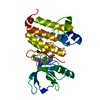

 SPODOPTERA FRUGIPERDA (fall armyworm)
 Mass: 493.599 Da / Num. of mol.: 1 / Source method: obtained synthetically / Formula: C30H31N5O2
 Mass: 18.015 Da / Num. of mol.: 91 / Source method: isolated from a natural source / Formula: H2O
 Sample preparation
 / Beamline: 32-ID / Wavelength: 1.00036
  Processing