 Movie
Movie Controller


+ Open data

- Basic information
| Entry | Database: PDB / ID: 3dae | ||||||
|---|---|---|---|---|---|---|---|
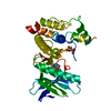
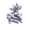
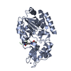
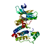
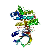
| Title | Crystal structure of phosphorylated SNF1 kinase domain | ||||||
 Components Components | Carbon catabolite-derepressing protein kinase | ||||||
 Keywords Keywords | TRANSFERASE / kinase / AMPK / snf1 / ATP-binding / Carbohydrate metabolism / Membrane / Nucleotide-binding / Nucleus / Phosphoprotein / Serine/threonine-protein kinase | ||||||
| Function / homology |  Function and homology information Function and homology informationfungal-type cell wall assembly / positive regulation of pseudohyphal growth / AMPK inhibits chREBP transcriptional activation activity / Energy dependent regulation of mTOR by LKB1-AMPK / regulation of cellular response to glucose starvation / single-species surface biofilm formation / positive regulation of filamentous growth of a population of unicellular organisms in response to starvation / regulation of invasive growth in response to glucose limitation / cellular bud neck septin ring / Carnitine shuttle ...fungal-type cell wall assembly / positive regulation of pseudohyphal growth / AMPK inhibits chREBP transcriptional activation activity / Energy dependent regulation of mTOR by LKB1-AMPK / regulation of cellular response to glucose starvation / single-species surface biofilm formation / positive regulation of filamentous growth of a population of unicellular organisms in response to starvation / regulation of invasive growth in response to glucose limitation / cellular bud neck septin ring / Carnitine shuttle / negative regulation of inositol phosphate biosynthetic process / invasive growth in response to glucose limitation / Macroautophagy / AMP-activated protein kinase activity / filamentous growth / nucleotide-activated protein kinase complex / vacuolar membrane / cellular response to stress / nuclear envelope lumen / establishment of mitotic spindle orientation / positive regulation of macroautophagy / response to unfolded protein / cellular response to glucose starvation / guanyl-nucleotide exchange factor activity / positive regulation of gluconeogenesis / molecular function activator activity / response to endoplasmic reticulum stress / nuclear membrane / protein kinase activity / non-specific serine/threonine protein kinase / negative regulation of translation / protein serine kinase activity / protein serine/threonine kinase activity / mitochondrion / ATP binding / identical protein binding / nucleus / cytoplasm Similarity search - Function | ||||||
| Biological species |  | ||||||
| Method |  X-RAY DIFFRACTION / SYNCHROTRON / MOLECULAR REPLACEMENT / Resolution: 2.899 Å X-RAY DIFFRACTION / SYNCHROTRON / MOLECULAR REPLACEMENT / Resolution: 2.899 Å | ||||||
 Authors Authors | Zheng, L.-S. / Chen, L. / Jiao, Z.-H. / Wu, J.-W. | ||||||
 Citation Citation | Journal: Nature / Year: 2009 Title: Structural insight into the autoinhibition mechanism of AMP-activated protein kinase Authors: Chen, L. / Jiao, Z.-H. / Zheng, L.-S. / Zhang, Y.-Y. / Xie, S.-T. / Wang, Z.-X. / Wu, J.-W. | ||||||
| History |
|
- Structure visualization
| Structure viewer | Molecule: MolmilJmol/JSmol |
|---|
- Downloads & links
-Download
| PDBx/mmCIF format | 3dae.cif.gz | 116 KB | Display | PDBx/mmCIF format |
|---|---|---|---|---|
| PDB format | pdb3dae.ent.gz | 89.1 KB | Display | PDB format |
| PDBx/mmJSON format | 3dae.json.gz | Tree view | PDBx/mmJSON format | |
| Others |  Other downloads Other downloads |
-Validation report
| Arichive directory | https://data.pdbj.org/pub/pdb/validation_reports/da/3daeftp://data.pdbj.org/pub/pdb/validation_reports/da/3dae | HTTPS FTP |
|---|
-Related structure data
| Related structure data | 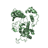 3h4jC  2eue S: Starting model for refinement C: citing same article ( |
|---|---|
| Similar structure data |


-Links
 PDBj
PDBj
- Assembly
| Deposited unit | 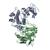
| ||||||||
|---|---|---|---|---|---|---|---|---|---|
| 1 | 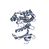
| ||||||||
| 2 | 
| ||||||||
| 3 |
| ||||||||
| Unit cell |
| ||||||||
| Details | THE DEPOSITORS THINK THE SOFTWARE PREDICTED QUATERNARY STRUCTURE IS NOT CONSISTENT WITH BIOCHEMICAL DATA AND MONOMER IS THE BIOLOGICAL UNIT. THE DIMER FORMATION MAY BE DUE TO CRYSTAL PACKING. |
-Components
| #1: Protein | Mass: 32436.666 Da / Num. of mol.: 2 / Fragment: Kinase Domain, UNP residue 41-315 Source method: isolated from a genetically manipulated source Source: (gene. exp.) Gene: SNF1 / Plasmid: pET21b / Production host:  References: UniProt: P06782, non-specific serine/threonine protein kinase #2: Water | ChemComp-HOH / |  Mass: 18.015 Da / Num. of mol.: 187 / Source method: isolated from a natural source / Formula: H2O Mass: 18.015 Da / Num. of mol.: 187 / Source method: isolated from a natural source / Formula: H2O |
|---|
-Experimental details
-Experiment
| Experiment | Method: X-RAY DIFFRACTION / Number of used crystals: 1 |
|---|
- Sample preparation
| Crystal | Density Matthews: 2.35 Å3/Da / Density % sol: 47.65 % |
|---|---|
| Crystal grow | Temperature: 298 K / Method: vapor diffusion, hanging drop / pH: 8.5 Details: Tris pH 8.5, 3.2-3.6M NH4Ac, vapor diffusion, hanging drop, temperature 298K |
-Data collection
| Diffraction | Mean temperature: 150 K |
|---|---|
| Diffraction source | Source: SYNCHROTRON / Site: BSRF  / Beamline: 3W1A / Wavelength: 1.00001 Å / Beamline: 3W1A / Wavelength: 1.00001 Å |
| Detector | Type: MAR CCD 165 mm / Detector: CCD / Date: Jun 24, 2007 |
| Radiation | Monochromator: double crystal monochromator / Protocol: SINGLE WAVELENGTH / Monochromatic (M) / Laue (L): M / Scattering type: x-ray |
| Radiation wavelength | Wavelength: 1.00001 Å / Relative weight: 1 |
| Reflection | Resolution: 2.899→47.2 Å / Num. all: 14122 / Num. obs: 14122 / % possible obs: 100 % / Observed criterion σ(F): 0 / Observed criterion σ(I): 0 / Redundancy: 7.4 % / Biso Wilson estimate: 50.35 Å2 / Rmerge(I) obs: 0.087 / Net I/σ(I): 20.7 / Num. measured all: 102012 |
| Reflection shell | Resolution: 2.899→3 Å / Redundancy: 7.2 % / Rmerge(I) obs: 0.382 / Mean I/σ(I) obs: 4.29 / Num. unique all: 1369 / % possible all: 100 |
- Processing
| Software |
| ||||||||||||||||||||||||||||||||||||
|---|---|---|---|---|---|---|---|---|---|---|---|---|---|---|---|---|---|---|---|---|---|---|---|---|---|---|---|---|---|---|---|---|---|---|---|---|---|
| Refinement | Method to determine structure: MOLECULAR REPLACEMENT Starting model: PDB ENTRY 2EUE 2eue Resolution: 2.899→47.192 Å / FOM work R set: 0.792 / σ(F): 1.36 / Stereochemistry target values: ML
| ||||||||||||||||||||||||||||||||||||
| Solvent computation | Shrinkage radii: 0.9 Å / VDW probe radii: 1.11 Å / Solvent model: FLAT BULK SOLVENT MODEL / Bsol: 30.136 Å2 / ksol: 0.317 e/Å3 | ||||||||||||||||||||||||||||||||||||
| Displacement parameters | Biso max: 8.63 Å2 / Biso mean: 53.73 Å2 / Biso min: 205.13 Å2
| ||||||||||||||||||||||||||||||||||||
| Refinement step | Cycle: LAST / Resolution: 2.899→47.192 Å
| ||||||||||||||||||||||||||||||||||||
| Refine LS restraints |
| ||||||||||||||||||||||||||||||||||||
| LS refinement shell | Refine-ID: X-RAY DIFFRACTION / Total num. of bins used: 5 / % reflection obs: 100 %
|
