 Movie
Movie Controller


+ Open data

- Basic information
| Entry | Database: PDB / ID: 5zgc | ||||||
|---|---|---|---|---|---|---|---|
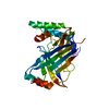
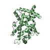
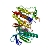
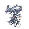
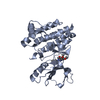
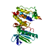
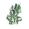
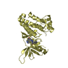
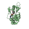
| Title | Crystal Structure of SIRT3 in complex with H4K16bhb peptide | ||||||
 Components Components |
| ||||||
 Keywords Keywords | HYDROLASE / Rossmann fold | ||||||
| Function / homology |  Function and homology information Function and homology informationpositive regulation of catalase activity / positive regulation of superoxide dismutase activity / NAD-dependent protein lysine delactylase activity / positive regulation of ceramide biosynthetic process / peptidyl-lysine deacetylation / Maturation of TCA enzymes and regulation of TCA cycle / protein acetyllysine N-acetyltransferase / protein deacetylation / NAD-dependent protein lysine deacetylase activity / histone deacetylase activity, NAD-dependent ...positive regulation of catalase activity / positive regulation of superoxide dismutase activity / NAD-dependent protein lysine delactylase activity / positive regulation of ceramide biosynthetic process / peptidyl-lysine deacetylation / Maturation of TCA enzymes and regulation of TCA cycle / protein acetyllysine N-acetyltransferase / protein deacetylation / NAD-dependent protein lysine deacetylase activity / histone deacetylase activity, NAD-dependent / positive regulation of oxidative phosphorylation / Regulation of FOXO transcriptional activity by acetylation / protein lysine deacetylase activity / cellular response to stress / negative regulation of reactive oxygen species metabolic process / NAD+ binding / Mitochondrial unfolded protein response (UPRmt) / FOXO-mediated transcription of oxidative stress, metabolic and neuronal genes / negative regulation of megakaryocyte differentiation / protein localization to CENP-A containing chromatin / Replacement of protamines by nucleosomes in the male pronucleus / CENP-A containing nucleosome / Packaging Of Telomere Ends / Transferases; Acyltransferases; Transferring groups other than aminoacyl groups / Recognition and association of DNA glycosylase with site containing an affected purine / Cleavage of the damaged purine / Deposition of new CENPA-containing nucleosomes at the centromere / telomere organization / Recognition and association of DNA glycosylase with site containing an affected pyrimidine / Cleavage of the damaged pyrimidine / RNA Polymerase I Promoter Opening / Inhibition of DNA recombination at telomere / aerobic respiration / Assembly of the ORC complex at the origin of replication / Meiotic synapsis / SUMOylation of chromatin organization proteins / Regulation of endogenous retroelements by the Human Silencing Hub (HUSH) complex / DNA methylation / Condensation of Prophase Chromosomes / Chromatin modifications during the maternal to zygotic transition (MZT) / SIRT1 negatively regulates rRNA expression / HCMV Late Events / ERCC6 (CSB) and EHMT2 (G9a) positively regulate rRNA expression / PRC2 methylates histones and DNA / Regulation of endogenous retroelements by KRAB-ZFP proteins / Defective pyroptosis / HDACs deacetylate histones / Regulation of endogenous retroelements by Piwi-interacting RNAs (piRNAs) / RNA Polymerase I Promoter Escape / Nonhomologous End-Joining (NHEJ) / Transcriptional activation of mitochondrial biogenesis / Transcriptional regulation by small RNAs / HDMs demethylate histones / Formation of the beta-catenin:TCF transactivating complex / Activated PKN1 stimulates transcription of AR (androgen receptor) regulated genes KLK2 and KLK3 / RUNX1 regulates genes involved in megakaryocyte differentiation and platelet function / negative regulation of ERK1 and ERK2 cascade / NoRC negatively regulates rRNA expression / Negative Regulation of CDH1 Gene Transcription / G2/M DNA damage checkpoint / PKMTs methylate histone lysines / B-WICH complex positively regulates rRNA expression / DNA Damage/Telomere Stress Induced Senescence / Meiotic recombination / Pre-NOTCH Transcription and Translation / Activation of anterior HOX genes in hindbrain development during early embryogenesis / Transcriptional regulation of granulopoiesis / RMTs methylate histone arginines / HCMV Early Events / positive regulation of insulin secretion / structural constituent of chromatin / nucleosome / nucleosome assembly / HATs acetylate histones / Recruitment and ATM-mediated phosphorylation of repair and signaling proteins at DNA double strand breaks / MLL4 and MLL3 complexes regulate expression of PPARG target genes in adipogenesis and hepatic steatosis / chromatin organization / RUNX1 regulates transcription of genes involved in differentiation of HSCs / Processing of DNA double-strand break ends / Senescence-Associated Secretory Phenotype (SASP) / Oxidative Stress Induced Senescence / Estrogen-dependent gene expression / sequence-specific DNA binding / chromosome, telomeric region / mitochondrial matrix / Amyloid fiber formation / protein heterodimerization activity / enzyme binding / protein-containing complex / mitochondrion / DNA binding / RNA binding / extracellular exosome / extracellular region / zinc ion binding / nucleoplasm / membrane / nucleus Similarity search - Function | ||||||
| Biological species |  Homo sapiens (human) Homo sapiens (human) | ||||||
| Method |  X-RAY DIFFRACTION / SYNCHROTRON / MOLECULAR REPLACEMENT / Resolution: 2.9 Å X-RAY DIFFRACTION / SYNCHROTRON / MOLECULAR REPLACEMENT / Resolution: 2.9 Å | ||||||
 Authors Authors | Li, H. / Zhang, X. | ||||||
 Citation Citation | Journal: To be published Title: Crystal Structure of SIRT3 in complex with H4K16bhb peptide Authors: Li, H. / Zhang, X. | ||||||
| History |
|
- Structure visualization
| Structure viewer | Molecule: MolmilJmol/JSmol |
|---|
- Downloads & links
-Download
| PDBx/mmCIF format | 5zgc.cif.gz | 315.4 KB | Display | PDBx/mmCIF format |
|---|---|---|---|---|
| PDB format | pdb5zgc.ent.gz | 256.7 KB | Display | PDB format |
| PDBx/mmJSON format | 5zgc.json.gz | Tree view | PDBx/mmJSON format | |
| Others |  Other downloads Other downloads |
-Validation report
| Arichive directory | https://data.pdbj.org/pub/pdb/validation_reports/zg/5zgcftp://data.pdbj.org/pub/pdb/validation_reports/zg/5zgc | HTTPS FTP |
|---|
-Related structure data
| Similar structure data |
|---|

-Links
 PDBj
PDBj


- Assembly
| Deposited unit | 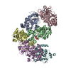
| ||||||||
|---|---|---|---|---|---|---|---|---|---|
| 1 | 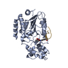
| ||||||||
| 2 | 
| ||||||||
| 3 | 
| ||||||||
| 4 | 
| ||||||||
| 5 | 
| ||||||||
| 6 | 
| ||||||||
| Unit cell |
|
-Components
| #1: Protein | Mass: 30495.225 Da / Num. of mol.: 6 / Fragment: UNP residues 121-394 Source method: isolated from a genetically manipulated source Source: (gene. exp.) Homo sapiens (human) / Gene: SIRT3 / Plasmid: pET28b / Production host:  References: UniProt: Q9NTG7, Hydrolases; Acting on carbon-nitrogen bonds, other than peptide bonds; In linear amides #2: Protein/peptide | Mass: 1284.535 Da / Num. of mol.: 6 / Source method: obtained synthetically / Source: (synth.) Homo sapiens (human) / References: UniProt: P62805*PLUS#3: Chemical | ChemComp-ZN /   Mass: 65.409 Da / Num. of mol.: 5 / Source method: obtained synthetically / Formula: Zn Mass: 65.409 Da / Num. of mol.: 5 / Source method: obtained synthetically / Formula: Zn#4: Water | ChemComp-HOH / |  Mass: 18.015 Da / Num. of mol.: 61 / Source method: isolated from a natural source / Formula: H2O Mass: 18.015 Da / Num. of mol.: 61 / Source method: isolated from a natural source / Formula: H2OHas protein modification | Y | |
|---|
-Experimental details
-Experiment
| Experiment | Method: X-RAY DIFFRACTION / Number of used crystals: 1 |
|---|
- Sample preparation
| Crystal | Density Matthews: 3.09 Å3/Da / Density % sol: 60.23 % |
|---|---|
| Crystal grow | Temperature: 291 K / Method: vapor diffusion, sitting drop / pH: 9 / Details: 0.1M Bicine pH 9.0, 10% PEG 6000 |
-Data collection
| Diffraction | Mean temperature: 100 K / Serial crystal experiment: N |
|---|---|
| Diffraction source | Source: SYNCHROTRON / Site: SSRF  / Beamline: BL17U1 / Wavelength: 0.9792 Å / Beamline: BL17U1 / Wavelength: 0.9792 Å |
| Detector | Type: DECTRIS EIGER X 16M / Detector: PIXEL / Date: Jan 25, 2018 / Details: mirrors |
| Radiation | Protocol: SINGLE WAVELENGTH / Monochromatic (M) / Laue (L): M / Scattering type: x-ray |
| Radiation wavelength | Wavelength: 0.9792 Å / Relative weight: 1 |
| Reflection | Resolution: 2.9→50 Å / Num. obs: 52894 / % possible obs: 99.6 % / Redundancy: 7.5 % / Biso Wilson estimate: 69.03 Å2 / Rpim(I) all: 0.039 / Net I/σ(I): 13.6 |
| Reflection shell | Resolution: 2.9→2.97 Å / Rpim(I) all: 0.47 |
- Processing
| Software |
| ||||||||||||||||||||||||||||||||||||||||||||||||||||||||||||||||||||||||||||||||||||||||||||||||||||||||||||||||||||||||||||||||||||||||||||
|---|---|---|---|---|---|---|---|---|---|---|---|---|---|---|---|---|---|---|---|---|---|---|---|---|---|---|---|---|---|---|---|---|---|---|---|---|---|---|---|---|---|---|---|---|---|---|---|---|---|---|---|---|---|---|---|---|---|---|---|---|---|---|---|---|---|---|---|---|---|---|---|---|---|---|---|---|---|---|---|---|---|---|---|---|---|---|---|---|---|---|---|---|---|---|---|---|---|---|---|---|---|---|---|---|---|---|---|---|---|---|---|---|---|---|---|---|---|---|---|---|---|---|---|---|---|---|---|---|---|---|---|---|---|---|---|---|---|---|---|---|---|
| Refinement | Method to determine structure: MOLECULAR REPLACEMENT / Resolution: 2.9→43.157 Å / SU ML: 0.45 / Cross valid method: THROUGHOUT / σ(F): 1.35 / Phase error: 29.63 / Stereochemistry target values: ML
| ||||||||||||||||||||||||||||||||||||||||||||||||||||||||||||||||||||||||||||||||||||||||||||||||||||||||||||||||||||||||||||||||||||||||||||
| Solvent computation | Shrinkage radii: 0.9 Å / VDW probe radii: 1.11 Å / Solvent model: FLAT BULK SOLVENT MODEL | ||||||||||||||||||||||||||||||||||||||||||||||||||||||||||||||||||||||||||||||||||||||||||||||||||||||||||||||||||||||||||||||||||||||||||||
| Displacement parameters | Biso max: 179.09 Å2 / Biso mean: 72.3898 Å2 / Biso min: 24.46 Å2 | ||||||||||||||||||||||||||||||||||||||||||||||||||||||||||||||||||||||||||||||||||||||||||||||||||||||||||||||||||||||||||||||||||||||||||||
| Refinement step | Cycle: final / Resolution: 2.9→43.157 Å
| ||||||||||||||||||||||||||||||||||||||||||||||||||||||||||||||||||||||||||||||||||||||||||||||||||||||||||||||||||||||||||||||||||||||||||||
| LS refinement shell | Refine-ID: X-RAY DIFFRACTION / Rfactor Rfree error: 0 / Total num. of bins used: 19
|
