[English] 日本語
Yorodumi
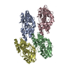
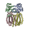
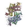
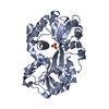
- PDB-2voz: Apo FutA2 from Synechocystis PCC6803 -

+
Open data


ID or keywords:

Loading...

-
Basic information

Entry
Database: PDB / ID: 2voz
TitleApo FutA2 from Synechocystis PCC6803
ComponentsPERIPLASMIC IRON-BINDING PROTEIN
KeywordsMETAL BINDING PROTEIN / FERRIC BINDING PROTEIN / METAL-BINDING PROTEIN / TAT / IRON / FUTA2 / SYNECHOCYSTIS
Function / homology
Function and homology information


plasma membrane-derived thylakoid membrane / outer membrane-bounded periplasmic space / iron ion transport / metal ion binding
Similarity search - Function
Ferric binding protein / Bacterial extracellular solute-binding protein / Bacterial extracellular solute-binding protein / Twin arginine translocation (Tat) signal profile. / Twin-arginine translocation pathway, signal sequence / Periplasmic binding protein-like II / D-Maltodextrin-Binding Protein; domain 2 / 3-Layer(aba) Sandwich / Alpha Beta
Similarity search - Domain/homology
Iron uptake protein A2
Similarity search - Component
Biological speciesSYNECHOCYSTIS SP. (bacteria)
MethodX-RAY DIFFRACTION / SYNCHROTRON / MOLECULAR REPLACEMENT / Resolution: 1.7 Å
AuthorsBadarau, A. / Firbank, S.J. / Banfield, M.J. / Dennison, C.
CitationJournal: J.Biol.Chem. / Year: 2008
Title: Futa2 is a Ferric Binding Protein from Synechocystis Pcc 6803.
Authors: Badarau, A. / Firbank, S.J. / Waldron, K.J. / Yanagisawa, S. / Robinson, N.J. / Banfield, M.J. / Dennison, C.
History
DepositionFeb 25, 2008Deposition site: PDBE / Processing site: PDBE
Revision 1.0Mar 25, 2008Provider: repository / Type: Initial release
Revision 1.1May 8, 2011Group: Version format compliance
Revision 1.2Jul 13, 2011Group: Version format compliance
Revision 1.3Dec 13, 2023Group: Data collection / Database references ...Data collection / Database references / Other / Refinement description
Category: chem_comp_atom / chem_comp_bond ...chem_comp_atom / chem_comp_bond / database_2 / pdbx_database_status / pdbx_initial_refinement_model
Item: _database_2.pdbx_DOI / _database_2.pdbx_database_accession / _pdbx_database_status.status_code_sf
Remark 700 SHEET THE SHEET STRUCTURE OF THIS MOLECULE IS BIFURCATED. IN ORDER TO REPRESENT THIS FEATURE IN ... SHEET THE SHEET STRUCTURE OF THIS MOLECULE IS BIFURCATED. IN ORDER TO REPRESENT THIS FEATURE IN THE SHEET RECORDS BELOW, TWO SHEETS ARE DEFINED.

-
Structure visualization

Structure viewerMolecule:
MolmilJmol/JSmol

Downloads & links

-
Assembly

Deposited unit
A: PERIPLASMIC IRON-BINDING PROTEIN
B: PERIPLASMIC IRON-BINDING PROTEIN
hetero molecules


Theoretical massNumber of molelcules
Total (without water)76,6795
Polymers76,3912
Non-polymers2883
Water14,394799
1
A: PERIPLASMIC IRON-BINDING PROTEIN
B: PERIPLASMIC IRON-BINDING PROTEIN
hetero molecules

A: PERIPLASMIC IRON-BINDING PROTEIN
B: PERIPLASMIC IRON-BINDING PROTEIN
hetero molecules


Theoretical massNumber of molelcules
Total (without water)153,35810
Polymers152,7824
Non-polymers5766
Water724
TypeNameSymmetry operationNumber
identity operation1_555x,y,z1
crystal symmetry operation2_555-x,-y,z1
Buried area8440 Å2
ΔGint-39.8 kcal/mol
Surface area52250 Å2
MethodPQS
2
A: PERIPLASMIC IRON-BINDING PROTEIN
hetero molecules


Theoretical massNumber of molelcules
Total (without water)38,3883
Polymers38,1961
Non-polymers1922
Water181
TypeNameSymmetry operationNumber
identity operation1_555x,y,z1
3
B: PERIPLASMIC IRON-BINDING PROTEIN
hetero molecules


Theoretical massNumber of molelcules
Total (without water)38,2922
Polymers38,1961
Non-polymers961
Water181
TypeNameSymmetry operationNumber
identity operation1_555x,y,z1
Unit cell
Length a, b, c (Å)91.080, 100.134, 66.523
Angle α, β, γ (deg.)90.00, 90.00, 90.00
Int Tables number18
Space group name H-MP21212

-
Components

#1: Protein PERIPLASMIC IRON-BINDING PROTEIN / FUTA2


Mass: 38195.508 Da / Num. of mol.: 2 / Source method: isolated from a natural source / Source: (natural) SYNECHOCYSTIS SP. (bacteria) / Strain: PCC 6803 / References: UniProt: Q55835
#2: Chemical ChemComp-SO4 / SULFATE ION / Sulfate


Mass: 96.063 Da / Num. of mol.: 3 / Source method: obtained synthetically / Formula: SO4
#3: Water ChemComp-HOH / water / Water


Mass: 18.015 Da / Num. of mol.: 799 / Source method: isolated from a natural source / Formula: H2O

-
Experimental details

-
Experiment

ExperimentMethod: X-RAY DIFFRACTION / Number of used crystals: 1

-
Sample preparation

CrystalDensity Matthews: 2.01 Å3/Da / Density % sol: 38 % / Description: NONE
Crystal growpH: 5.6
Details: 0.5M AMMONIUM SULPHATE, 1M LITHIUM SULPHATE, 0.1M TRI-SODIUM CITRATE PH 5.6

-
Data collection

DiffractionMean temperature: 100 K
Diffraction sourceSource: SYNCHROTRON / Site: SRS / Beamline: PX10.1 / Wavelength: 0.979
DetectorType: MARRESEARCH / Detector: CCD / Details: RH COATED COLLIMATING MIRROR
RadiationMonochromator: DOUBLE CRYSTAL SI(III) / Protocol: SINGLE WAVELENGTH / Monochromatic (M) / Laue (L): M / Scattering type: x-ray
Radiation wavelengthWavelength: 0.979 Å / Relative weight: 1
ReflectionResolution: 1.7→44.4 Å / Num. obs: 67656 / % possible obs: 100 % / Observed criterion σ(I): 0 / Redundancy: 5.6 % / Rmerge(I) obs: 0.06 / Net I/σ(I): 18.4
Reflection shellResolution: 1.7→1.79 Å / Redundancy: 5.6 % / Rmerge(I) obs: 0.28 / Mean I/σ(I) obs: 4.5 / % possible all: 100

-
Processing

Software
NameVersionClassification
REFMAC5.3.0034refinement
MOSFLMdata reduction
SCALAdata scaling
PHASERphasing
RefinementMethod to determine structure: MOLECULAR REPLACEMENT
Starting model: PDB ENTRY 1Q35
Resolution: 1.7→19.54 Å / Cor.coef. Fo:Fc: 0.959 / Cor.coef. Fo:Fc free: 0.946 / SU B: 1.957 / SU ML: 0.066 / Cross valid method: THROUGHOUT / ESU R: 0.105 / ESU R Free: 0.104 / Stereochemistry target values: MAXIMUM LIKELIHOOD / Details: HYDROGENS HAVE BEEN ADDED IN THE RIDING POSITIONS.
RfactorNum. reflection% reflectionSelection details
Rfree0.201 3385 5 %RANDOM
Rwork0.163 ---
obs0.165 64137 100 %-
Solvent computationIon probe radii: 0.8 Å / Shrinkage radii: 0.8 Å / VDW probe radii: 1.2 Å / Solvent model: MASK
Displacement parametersBiso mean: 15.57 Å2
Baniso -1Baniso -2Baniso -3
1-0 Å20 Å20 Å2
2--0 Å20 Å2
3---0 Å2
Refinement stepCycle: LAST / Resolution: 1.7→19.54 Å
ProteinNucleic acidLigandSolventTotal
Num. atoms4873 0 15 799 5687
Refine LS restraints
Refine-IDTypeDev idealDev ideal targetNumber
X-RAY DIFFRACTIONr_bond_refined_d0.0150.0225089
X-RAY DIFFRACTIONr_bond_other_d
X-RAY DIFFRACTIONr_angle_refined_deg1.5281.9536964
X-RAY DIFFRACTIONr_angle_other_deg
X-RAY DIFFRACTIONr_dihedral_angle_1_deg5.7335660
X-RAY DIFFRACTIONr_dihedral_angle_2_deg38.81424.023261
X-RAY DIFFRACTIONr_dihedral_angle_3_deg12.29315777
X-RAY DIFFRACTIONr_dihedral_angle_4_deg13.6071544
X-RAY DIFFRACTIONr_chiral_restr0.1210.2748
X-RAY DIFFRACTIONr_gen_planes_refined0.0070.024066
X-RAY DIFFRACTIONr_gen_planes_other
X-RAY DIFFRACTIONr_nbd_refined0.2060.22657
X-RAY DIFFRACTIONr_nbd_other
X-RAY DIFFRACTIONr_nbtor_refined0.3120.23560
X-RAY DIFFRACTIONr_nbtor_other
X-RAY DIFFRACTIONr_xyhbond_nbd_refined0.1540.2670
X-RAY DIFFRACTIONr_xyhbond_nbd_other
X-RAY DIFFRACTIONr_metal_ion_refined
X-RAY DIFFRACTIONr_metal_ion_other
X-RAY DIFFRACTIONr_symmetry_vdw_refined0.2120.252
X-RAY DIFFRACTIONr_symmetry_vdw_other
X-RAY DIFFRACTIONr_symmetry_hbond_refined0.1680.254
X-RAY DIFFRACTIONr_symmetry_hbond_other
X-RAY DIFFRACTIONr_symmetry_metal_ion_refined
X-RAY DIFFRACTIONr_symmetry_metal_ion_other
X-RAY DIFFRACTIONr_mcbond_it0.9371.53230
X-RAY DIFFRACTIONr_mcbond_other
X-RAY DIFFRACTIONr_mcangle_it1.47425105
X-RAY DIFFRACTIONr_mcangle_other
X-RAY DIFFRACTIONr_scbond_it2.40332093
X-RAY DIFFRACTIONr_scbond_other
X-RAY DIFFRACTIONr_scangle_it3.5524.51842
X-RAY DIFFRACTIONr_scangle_other
X-RAY DIFFRACTIONr_long_range_B_refined
X-RAY DIFFRACTIONr_long_range_B_other
X-RAY DIFFRACTIONr_rigid_bond_restr
X-RAY DIFFRACTIONr_sphericity_free
X-RAY DIFFRACTIONr_sphericity_bonded
LS refinement shellResolution: 1.7→1.74 Å / Total num. of bins used: 20 /
RfactorNum. reflection
Rfree0.32 218
Rwork0.206 4689

+
About Yorodumi

-
News

-
Feb 9, 2022. New format data for meta-information of EMDB entries

New format data for meta-information of EMDB entries

  • Version 3 of the EMDB header file is now the official format.
  • The previous official version 1.9 will be removed from the archive.

Related info.:EMDB header

External links:wwPDB to switch to version 3 of the EMDB data model

-
Aug 12, 2020. Covid-19 info

Covid-19 info

URL: https://pdbj.org/emnavi/covid19.php

New page: Covid-19 featured information page in EM Navigator.

Related info.:Covid-19 info / Mar 5, 2020. Novel coronavirus structure data

+
Mar 5, 2020. Novel coronavirus structure data

Novel coronavirus structure data

Related info.:Yorodumi Speices / Aug 12, 2020. Covid-19 info

External links:COVID-19 featured content - PDBj / Molecule of the Month (242):Coronavirus Proteases

+
Jan 31, 2019. EMDB accession codes are about to change! (news from PDBe EMDB page)

EMDB accession codes are about to change! (news from PDBe EMDB page)

  • The allocation of 4 digits for EMDB accession codes will soon come to an end. Whilst these codes will remain in use, new EMDB accession codes will include an additional digit and will expand incrementally as the available range of codes is exhausted. The current 4-digit format prefixed with “EMD-” (i.e. EMD-XXXX) will advance to a 5-digit format (i.e. EMD-XXXXX), and so on. It is currently estimated that the 4-digit codes will be depleted around Spring 2019, at which point the 5-digit format will come into force.
  • The EM Navigator/Yorodumi systems omit the EMD- prefix.

Related info.:Q: What is EMD? / ID/Accession-code notation in Yorodumi/EM Navigator

External links:EMDB Accession Codes are Changing Soon! / Contact to PDBj

+
Jul 12, 2017. Major update of PDB

Major update of PDB

  • wwPDB released updated PDB data conforming to the new PDBx/mmCIF dictionary.
  • This is a major update changing the version number from 4 to 5, and with Remediation, in which all the entries are updated.
  • In this update, many items about electron microscopy experimental information are reorganized (e.g. em_software).
  • Now, EM Navigator and Yorodumi are based on the updated data.

External links:wwPDB Remediation / Enriched Model Files Conforming to OneDep Data Standards Now Available in the PDB FTP Archive

-
Yorodumi

Thousand views of thousand structures

  • Yorodumi is a browser for structure data from EMDB, PDB, SASBDB, etc.
  • This page is also the successor to EM Navigator detail page, and also detail information page/front-end page for Omokage search.
  • The word "yorodu" (or yorozu) is an old Japanese word meaning "ten thousand". "mi" (miru) is to see.

Related info.:EMDB / PDB / SASBDB / Comparison of 3 databanks / Yorodumi Search / Aug 31, 2016. New EM Navigator & Yorodumi / Yorodumi Papers / Jmol/JSmol / Function and homology information / Changes in new EM Navigator and Yorodumi

Read more