 Movie
Movie Controller


+ Open data

- Basic information
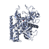
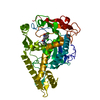
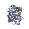
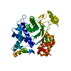
| Entry | Database: PDB / ID: 3h4j | ||||||
|---|---|---|---|---|---|---|---|
| Title | crystal structure of pombe AMPK KDAID fragment | ||||||
 Components Components | SNF1-like protein kinase ssp2 | ||||||
 Keywords Keywords | TRANSFERASE / AMPK / kinase / ATP-binding / Nucleotide-binding / Phosphoprotein / Serine/threonine-protein kinase | ||||||
| Function / homology |  Function and homology information Function and homology informationpositive regulation of cell cycle switching, mitotic to meiotic cell cycle / AMPK inhibits chREBP transcriptional activation activity / Carnitine shuttle / : / Energy dependent regulation of mTOR by LKB1-AMPK / TP53 Regulates Metabolic Genes / Macroautophagy / mitotic spindle pole body / SREBP signaling pathway / CAMKK-AMPK signaling cascade ...positive regulation of cell cycle switching, mitotic to meiotic cell cycle / AMPK inhibits chREBP transcriptional activation activity / Carnitine shuttle / : / Energy dependent regulation of mTOR by LKB1-AMPK / TP53 Regulates Metabolic Genes / Macroautophagy / mitotic spindle pole body / SREBP signaling pathway / CAMKK-AMPK signaling cascade / nucleotide-activated protein kinase complex / negative regulation of cytoplasmic translation / negative regulation of TORC1 signaling / non-specific serine/threonine protein kinase / protein serine kinase activity / protein serine/threonine kinase activity / positive regulation of transcription by RNA polymerase II / ATP binding / nucleus / cytoplasm / cytosol Similarity search - Function | ||||||
| Biological species |  | ||||||
| Method |  X-RAY DIFFRACTION / SYNCHROTRON / MOLECULAR REPLACEMENT / Resolution: 2.8 Å X-RAY DIFFRACTION / SYNCHROTRON / MOLECULAR REPLACEMENT / Resolution: 2.8 Å | ||||||
 Authors Authors | Chen, L. / Jiao, Z.-H. / Zheng, L.-S. / Zhang, Y.-Y. / Xie, S.-T. / Wang, Z.-X. / Wu, J.-W. | ||||||
 Citation Citation | Journal: Nature / Year: 2009 Title: Structural insight into the autoinhibition mechanism of AMP-activated protein kinase Authors: Chen, L. / Jiao, Z.-H. / Zheng, L.-S. / Zhang, Y.-Y. / Xie, S.-T. / Wang, Z.-X. / Wu, J.-W. | ||||||
| History |
|
- Structure visualization
| Structure viewer | Molecule: MolmilJmol/JSmol |
|---|
- Downloads & links
-Download
| PDBx/mmCIF format | 3h4j.cif.gz | 278.5 KB | Display | PDBx/mmCIF format |
|---|---|---|---|---|
| PDB format | pdb3h4j.ent.gz | 227.1 KB | Display | PDB format |
| PDBx/mmJSON format | 3h4j.json.gz | Tree view | PDBx/mmJSON format | |
| Others |  Other downloads Other downloads |
-Validation report
| Arichive directory | https://data.pdbj.org/pub/pdb/validation_reports/h4/3h4jftp://data.pdbj.org/pub/pdb/validation_reports/h4/3h4j | HTTPS FTP |
|---|
-Related structure data
| Related structure data |  3daeC  2eue S: Starting model for refinement C: citing same article ( |
|---|---|
| Similar structure data |


-Links
 PDBj
PDBj- Assembly
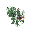
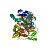
| Deposited unit | 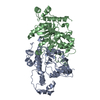
| ||||||||
|---|---|---|---|---|---|---|---|---|---|
| 1 | 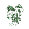
| ||||||||
| 2 | 
| ||||||||
| 3 |
| ||||||||
| Unit cell |
|
-Components
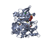
| #1: Protein | Mass: 38987.988 Da / Num. of mol.: 2 / Fragment: UNP residues 25-351 Source method: isolated from a genetically manipulated source Source: (gene. exp.) Strain: SPQ-01 / Gene: ssp2 / Plasmid: pET28b / Production host:  References: UniProt: O74536, non-specific serine/threonine protein kinase #2: Water | ChemComp-HOH / |  Mass: 18.015 Da / Num. of mol.: 104 / Source method: isolated from a natural source / Formula: H2O Mass: 18.015 Da / Num. of mol.: 104 / Source method: isolated from a natural source / Formula: H2O |
|---|
-Experimental details
-Experiment
| Experiment | Method: X-RAY DIFFRACTION / Number of used crystals: 1 |
|---|
- Sample preparation
| Crystal | Density Matthews: 3.29 Å3/Da / Density % sol: 62.62 % |
|---|---|
| Crystal grow | Temperature: 294 K / Method: vapor diffusion, hanging drop / pH: 5.6 Details: 0.1M sodium citrate, pH 5.6, 1.2M ammonium sulphate, VAPOR DIFFUSION, HANGING DROP, temperature 294K |
-Data collection
| Diffraction | Mean temperature: 100 K |
|---|---|
| Diffraction source | Source: SYNCHROTRON / Site: Photon Factory  / Beamline: AR-NW12A / Wavelength: 1 Å / Beamline: AR-NW12A / Wavelength: 1 Å |
| Detector | Type: ADSC QUANTUM 210r / Detector: CCD / Date: Dec 12, 2007 |
| Radiation | Protocol: SINGLE WAVELENGTH / Monochromatic (M) / Laue (L): M / Scattering type: x-ray |
| Radiation wavelength | Wavelength: 1 Å / Relative weight: 1 |
| Reflection | Resolution: 2.7→50 Å / Num. obs: 27824 / % possible obs: 99.8 % / Observed criterion σ(F): 0 / Observed criterion σ(I): 0 / Biso Wilson estimate: 71.11 Å2 |
| Reflection shell | Resolution: 2.7→2.8 Å / % possible all: 99.3 |
- Processing
| Software |
| ||||||||||||||||||||||||||||||||||||||||||||||||||||||||||||||||||||||
|---|---|---|---|---|---|---|---|---|---|---|---|---|---|---|---|---|---|---|---|---|---|---|---|---|---|---|---|---|---|---|---|---|---|---|---|---|---|---|---|---|---|---|---|---|---|---|---|---|---|---|---|---|---|---|---|---|---|---|---|---|---|---|---|---|---|---|---|---|---|---|---|
| Refinement | Method to determine structure: MOLECULAR REPLACEMENT Starting model: PDB ENTRY 2EUE 2eue Resolution: 2.8→39.262 Å / Occupancy max: 1 / Occupancy min: 1 / FOM work R set: 0.759 / SU ML: 0.39 / σ(F): 1.33 / Stereochemistry target values: ML
| ||||||||||||||||||||||||||||||||||||||||||||||||||||||||||||||||||||||
| Solvent computation | Shrinkage radii: 0.9 Å / VDW probe radii: 1.11 Å / Solvent model: FLAT BULK SOLVENT MODEL / Bsol: 81.756 Å2 / ksol: 0.313 e/Å3 | ||||||||||||||||||||||||||||||||||||||||||||||||||||||||||||||||||||||
| Displacement parameters | Biso max: 294.06 Å2 / Biso mean: 102.318 Å2 / Biso min: 31.81 Å2
| ||||||||||||||||||||||||||||||||||||||||||||||||||||||||||||||||||||||
| Refinement step | Cycle: LAST / Resolution: 2.8→39.262 Å
| ||||||||||||||||||||||||||||||||||||||||||||||||||||||||||||||||||||||
| Refine LS restraints |
| ||||||||||||||||||||||||||||||||||||||||||||||||||||||||||||||||||||||
| LS refinement shell | Refine-ID: X-RAY DIFFRACTION / Total num. of bins used: 9
|
