 Movie
Movie Controller

[English] 日本語
 Yorodumi
- PDB-6xce: Structure of the C. botulinum neurotoxin serotype A light chain p... -
+ Open data

- Basic information
| Entry | Database: PDB / ID: 6xce | ||||||
|---|---|---|---|---|---|---|---|
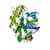
| Title | Structure of the C. botulinum neurotoxin serotype A light chain protease in complex with covalent inhibitor 53 | ||||||
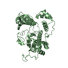
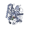
 Components Components | Botulinum neurotoxin type A | ||||||
 Keywords Keywords | TOXIN/INHIBITOR / covalent inhibitor / hydroxamate / TOXIN / TOXIN-INHIBITOR complex | ||||||
| Function / homology |  Function and homology information Function and homology informationsymbiont-mediated suppression of host exocytosis / Toxicity of botulinum toxin type A (botA) / ganglioside GT1b binding / bontoxilysin / host cell presynaptic membrane / host cell cytoplasmic vesicle / host cell cytosol / transmembrane protein transporter activity / metalloendopeptidase activity / toxin activity ...symbiont-mediated suppression of host exocytosis / Toxicity of botulinum toxin type A (botA) / ganglioside GT1b binding / bontoxilysin / host cell presynaptic membrane / host cell cytoplasmic vesicle / host cell cytosol / transmembrane protein transporter activity / metalloendopeptidase activity / toxin activity / proteolysis / extracellular region / zinc ion binding Similarity search - Function | ||||||
| Biological species |   Clostridium botulinum (bacteria) Clostridium botulinum (bacteria) | ||||||
| Method |  X-RAY DIFFRACTION / MOLECULAR REPLACEMENT / Resolution: 2.5 Å X-RAY DIFFRACTION / MOLECULAR REPLACEMENT / Resolution: 2.5 Å | ||||||
 Authors Authors | Tararina, M.A. / Allen, K.N. | ||||||
| Funding support |  United States, 1items United States, 1items
| ||||||
 Citation Citation | Journal: J.Med.Chem. / Year: 2020 Title: Catch and Anchor Approach To Combat Both Toxicity and Longevity of Botulinum Toxin A. Authors: Lin, L. / Olson, M.E. / Sugane, T. / Turner, L.D. / Tararina, M.A. / Nielsen, A.L. / Kurbanov, E.K. / Pellett, S. / Johnson, E.A. / Cohen, S.M. / Allen, K.N. / Janda, K.D. | ||||||
| History |
|
- Structure visualization
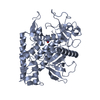
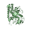
| Structure viewer | Molecule: MolmilJmol/JSmol |
|---|
- Downloads & links
-Download
| PDBx/mmCIF format | 6xce.cif.gz | 123.4 KB | Display | PDBx/mmCIF format |
|---|---|---|---|---|
| PDB format | pdb6xce.ent.gz | 75.4 KB | Display | PDB format |
| PDBx/mmJSON format | 6xce.json.gz | Tree view | PDBx/mmJSON format | |
| Others |  Other downloads Other downloads |
-Validation report
| Arichive directory | https://data.pdbj.org/pub/pdb/validation_reports/xc/6xceftp://data.pdbj.org/pub/pdb/validation_reports/xc/6xce | HTTPS FTP |
|---|
-Related structure data
| Related structure data |  6xcbC  6xccC 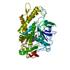 6xcdC  6xcfC  3bonS S: Starting model for refinement C: citing same article ( |
|---|---|
| Similar structure data |

-Links
 PDBj
PDBj
- Assembly
| Deposited unit | 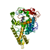
| ||||||||||||
|---|---|---|---|---|---|---|---|---|---|---|---|---|---|
| 1 |
| ||||||||||||
| Unit cell |
|
-Components
| #1: Protein | Mass: 50454.875 Da / Num. of mol.: 1 Source method: isolated from a genetically manipulated source Source: (gene. exp.) Clostridium botulinum (bacteria) / Gene: botA, atx, bonT / Production host: |
|---|---|
| #2: Chemical | ChemComp-UZM / (  Mass: 365.275 Da / Num. of mol.: 1 / Source method: obtained synthetically / Formula: C14H18Cl2N2O3S / Feature type: SUBJECT OF INVESTIGATION Mass: 365.275 Da / Num. of mol.: 1 / Source method: obtained synthetically / Formula: C14H18Cl2N2O3S / Feature type: SUBJECT OF INVESTIGATION |
| #3: Water | ChemComp-HOH /  Mass: 18.015 Da / Num. of mol.: 55 / Source method: isolated from a natural source / Formula: H2O Mass: 18.015 Da / Num. of mol.: 55 / Source method: isolated from a natural source / Formula: H2O |
| Has ligand of interest | Y |
| Has protein modification | Y |
-Experimental details
-Experiment
| Experiment | Method: X-RAY DIFFRACTION / Number of used crystals: 1 |
|---|
- Sample preparation
| Crystal | Density Matthews: 2.12 Å3/Da / Density % sol: 42.11 % |
|---|---|
| Crystal grow | Temperature: 290 K / Method: vapor diffusion, hanging drop / pH: 7 Details: 0.3 - 0.45 M ammonium tartrate dibasic, 15 - 25% PEG 3350 |
-Data collection
| Diffraction | Mean temperature: 100 K / Serial crystal experiment: N |
|---|---|
| Diffraction source | Source: SEALED TUBE / Type: BRUKER IMUS MICROFOCUS / Wavelength: 1.54178 Å |
| Detector | Type: APEX II CCD / Detector: CCD / Date: Oct 17, 2018 |
| Radiation | Protocol: SINGLE WAVELENGTH / Monochromatic (M) / Laue (L): M / Scattering type: x-ray |
| Radiation wavelength | Wavelength: 1.54178 Å / Relative weight: 1 |
| Reflection | Resolution: 2.5→29.55 Å / Num. obs: 14436 / % possible obs: 95.3 % / Redundancy: 6.7 % / Biso Wilson estimate: 30.05 Å2 / CC1/2: 0.96 / Rmerge(I) obs: 0.2 / Net I/σ(I): 7.1 |
| Reflection shell | Resolution: 2.5→2.59 Å / Redundancy: 4 % / Rmerge(I) obs: 0.578 / Mean I/σ(I) obs: 1.6 / Num. unique obs: 1233 / CC1/2: 0.74 / % possible all: 83.1 |
- Processing
| Software |
| |||||||||||||||||||||||||||||||||||||||||||||||||||||||||||||||||||||||||||||
|---|---|---|---|---|---|---|---|---|---|---|---|---|---|---|---|---|---|---|---|---|---|---|---|---|---|---|---|---|---|---|---|---|---|---|---|---|---|---|---|---|---|---|---|---|---|---|---|---|---|---|---|---|---|---|---|---|---|---|---|---|---|---|---|---|---|---|---|---|---|---|---|---|---|---|---|---|---|---|
| Refinement | Method to determine structure: MOLECULAR REPLACEMENT Starting model: 3BON Resolution: 2.5→29.55 Å / SU ML: 0.3005 / Cross valid method: FREE R-VALUE / σ(F): 1.33 / Phase error: 29.9644 / Stereochemistry target values: CDL v1.2
| |||||||||||||||||||||||||||||||||||||||||||||||||||||||||||||||||||||||||||||
| Solvent computation | Shrinkage radii: 0.9 Å / VDW probe radii: 1.11 Å / Solvent model: FLAT BULK SOLVENT MODEL | |||||||||||||||||||||||||||||||||||||||||||||||||||||||||||||||||||||||||||||
| Displacement parameters | Biso mean: 34.63 Å2 | |||||||||||||||||||||||||||||||||||||||||||||||||||||||||||||||||||||||||||||
| Refinement step | Cycle: LAST / Resolution: 2.5→29.55 Å
| |||||||||||||||||||||||||||||||||||||||||||||||||||||||||||||||||||||||||||||
| Refine LS restraints |
| |||||||||||||||||||||||||||||||||||||||||||||||||||||||||||||||||||||||||||||
| LS refinement shell |
|
