 Movie
Movie Controller

[English] 日本語
 Yorodumi
- PDB-1fgi: CRYSTAL STRUCTURE OF THE TYROSINE KINASE DOMAIN OF FIBROBLAST GRO... -
+ Open data

- Basic information
| Entry | Database: PDB / ID: 1fgi | ||||||
|---|---|---|---|---|---|---|---|
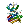
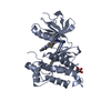
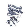
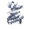
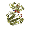
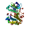
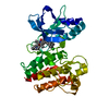
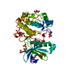
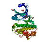
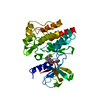
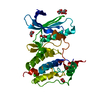
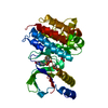
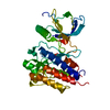
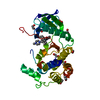
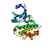
| Title | CRYSTAL STRUCTURE OF THE TYROSINE KINASE DOMAIN OF FIBROBLAST GROWTH FACTOR RECEPTOR 1 IN COMPLEX WITH SU5402 INHIBITOR | ||||||
 Components Components | FGF RECEPTOR 1 | ||||||
 Keywords Keywords | PROTEIN KINASE / TRANSFERASE / TYROSINE-PROTEIN KINASE / ATP-BINDING / PHOSPHORYLATION / INHIBITOR | ||||||
| Function / homology |  Function and homology information Function and homology informationSignaling by FGFR1 amplification mutants / negative regulation of fibroblast growth factor production / positive regulation of mitotic cell cycle DNA replication / regulation of extrinsic apoptotic signaling pathway in absence of ligand / diphosphate metabolic process / Signaling by plasma membrane FGFR1 fusions / FGFR1c and Klotho ligand binding and activation / regulation of lateral mesodermal cell fate specification / positive regulation of MAPKKK cascade by fibroblast growth factor receptor signaling pathway / regulation of phosphate transport ...Signaling by FGFR1 amplification mutants / negative regulation of fibroblast growth factor production / positive regulation of mitotic cell cycle DNA replication / regulation of extrinsic apoptotic signaling pathway in absence of ligand / diphosphate metabolic process / Signaling by plasma membrane FGFR1 fusions / FGFR1c and Klotho ligand binding and activation / regulation of lateral mesodermal cell fate specification / positive regulation of MAPKKK cascade by fibroblast growth factor receptor signaling pathway / regulation of phosphate transport / vitamin D3 metabolic process / cementum mineralization / regulation of branching involved in salivary gland morphogenesis by mesenchymal-epithelial signaling / response to sodium phosphate / Epithelial-Mesenchymal Transition (EMT) during gastrulation / fibroblast growth factor receptor signaling pathway involved in orbitofrontal cortex development / ventricular zone neuroblast division / mesenchymal cell proliferation / positive regulation of phospholipase activity / receptor-receptor interaction / chordate embryonic development / positive regulation of parathyroid hormone secretion / auditory receptor cell development / paraxial mesoderm development / regulation of postsynaptic density assembly / FGFR1b ligand binding and activation / organ induction / Signaling by activated point mutants of FGFR1 / FGFR1c ligand binding and activation / Downstream signaling of activated FGFR1 / fibroblast growth factor receptor activity / Phospholipase C-mediated cascade: FGFR1 / branching involved in salivary gland morphogenesis / lung-associated mesenchyme development / cell projection assembly / outer ear morphogenesis / embryonic limb morphogenesis / positive regulation of vascular endothelial cell proliferation / positive regulation of endothelial cell chemotaxis / ureteric bud development / positive regulation of mesenchymal cell proliferation / skeletal system morphogenesis / inner ear morphogenesis / middle ear morphogenesis / Formation of paraxial mesoderm / positive regulation of stem cell proliferation / PI-3K cascade:FGFR1 / midbrain development / positive regulation of MAP kinase activity / phosphatidylinositol-mediated signaling / regulation of cell differentiation / fibroblast growth factor binding / fibroblast growth factor receptor signaling pathway / PI3K Cascade / epithelial to mesenchymal transition / positive regulation of blood vessel endothelial cell migration / chondrocyte differentiation / cardiac muscle cell proliferation / positive regulation of cardiac muscle cell proliferation / calcium ion homeostasis / SHC-mediated cascade:FGFR1 / peptidyl-tyrosine phosphorylation / FRS-mediated FGFR1 signaling / cell maturation / cellular response to fibroblast growth factor stimulus / positive regulation of neuron differentiation / Signaling by FGFR1 in disease / NCAM signaling for neurite out-growth / SH2 domain binding / stem cell proliferation / stem cell differentiation / Signal transduction by L1 / skeletal system development / positive regulation of cell differentiation / Negative regulation of FGFR1 signaling / sensory perception of sound / receptor protein-tyrosine kinase / positive regulation of neuron projection development / neuron migration / Constitutive Signaling by Aberrant PI3K in Cancer / neuron projection development / protein autophosphorylation / PIP3 activates AKT signaling / MAPK cascade / cell migration / heparin binding / PI5P, PP2A and IER3 Regulate PI3K/AKT Signaling / RAF/MAP kinase cascade / protein tyrosine kinase activity / angiogenesis / cytoplasmic vesicle / gene expression / in utero embryonic development / protein phosphorylation / positive regulation of MAPK cascade / positive regulation of phosphatidylinositol 3-kinase/protein kinase B signal transduction / signaling receptor complex / postsynapse / positive regulation of cell population proliferation / glutamatergic synapse Similarity search - Function | ||||||
| Biological species |  Homo sapiens (human) Homo sapiens (human) | ||||||
| Method |  X-RAY DIFFRACTION / DIFFERENCE FOURIER / Resolution: 2.5 Å X-RAY DIFFRACTION / DIFFERENCE FOURIER / Resolution: 2.5 Å | ||||||
 Authors Authors | Mohammadi, M. / Schlessinger, J. / Hubbard, S.R. | ||||||
 Citation Citation | Journal: Science / Year: 1997 Title: Structures of the tyrosine kinase domain of fibroblast growth factor receptor in complex with inhibitors. Authors: Mohammadi, M. / McMahon, G. / Sun, L. / Tang, C. / Hirth, P. / Yeh, B.K. / Hubbard, S.R. / Schlessinger, J. #1: Journal: Cell(Cambridge,Mass.) / Year: 1996Title: Structure of the Fgf Receptor Tyrosine Kinase Domain Reveals a Novel Autoinhibitory Mechanism Authors: Mohammadi, M. / Schlessinger, J. / Hubbard, S.R. | ||||||
| History |
|
- Structure visualization
| Structure viewer | Molecule: MolmilJmol/JSmol |
|---|
- Downloads & links
-Download
| PDBx/mmCIF format | 1fgi.cif.gz | 126.8 KB | Display | PDBx/mmCIF format |
|---|---|---|---|---|
| PDB format | pdb1fgi.ent.gz | 97 KB | Display | PDB format |
| PDBx/mmJSON format | 1fgi.json.gz | Tree view | PDBx/mmJSON format | |
| Others |  Other downloads Other downloads |
-Validation report
| Arichive directory | https://data.pdbj.org/pub/pdb/validation_reports/fg/1fgiftp://data.pdbj.org/pub/pdb/validation_reports/fg/1fgi | HTTPS FTP |
|---|
-Related structure data


-Links
 PDBj
PDBj


- Assembly
| Deposited unit | 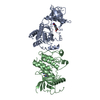
| ||||||||
|---|---|---|---|---|---|---|---|---|---|
| 1 | 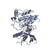
| ||||||||
| 2 | 
| ||||||||
| Unit cell |
| ||||||||
| Noncrystallographic symmetry (NCS) | NCS oper: (Code: given Matrix: (-0.9944, -0.09773, 0.04029), Vector: |
-Components
| #1: Protein | Mass: 35308.613 Da / Num. of mol.: 2 / Fragment: TYROSINE KINASE DOMAIN / Mutation: L457V, C488A, C584S Source method: isolated from a genetically manipulated source Source: (gene. exp.) Homo sapiens (human) / Cell line: SF9 / Cellular location: CYTOPLASM / Organelle: CYTOPLASM / Cell line (production host): SF9 / Production host:   Spodoptera frugiperda (fall armyworm) / References: UniProt: P11362, EC: 2.7.1.112 Spodoptera frugiperda (fall armyworm) / References: UniProt: P11362, EC: 2.7.1.112#2: Chemical |   Mass: 296.321 Da / Num. of mol.: 2 / Source method: obtained synthetically / Formula: C17H16N2O3 Mass: 296.321 Da / Num. of mol.: 2 / Source method: obtained synthetically / Formula: C17H16N2O3#3: Water | ChemComp-HOH / |  Mass: 18.015 Da / Num. of mol.: 229 / Source method: isolated from a natural source / Formula: H2O Mass: 18.015 Da / Num. of mol.: 229 / Source method: isolated from a natural source / Formula: H2O |
|---|
-Experimental details
-Experiment
| Experiment | Method: X-RAY DIFFRACTION / Number of used crystals: 1 |
|---|
- Sample preparation
| Crystal | Density Matthews: 2.68 Å3/Da / Density % sol: 54 % | ||||||||||||||||||||||||||||||||||||||||||||||||||||||
|---|---|---|---|---|---|---|---|---|---|---|---|---|---|---|---|---|---|---|---|---|---|---|---|---|---|---|---|---|---|---|---|---|---|---|---|---|---|---|---|---|---|---|---|---|---|---|---|---|---|---|---|---|---|---|---|
| Crystal grow | pH: 6.5 Details: 16% PEG 10000, 0.3 M (NH4)2SO4, 100 MM BIS-TRIS, PH 6.5, 5% ETHYLENE GLYCOL | ||||||||||||||||||||||||||||||||||||||||||||||||||||||
| Crystal grow | *PLUS Temperature: 4 ℃ / Method: vapor diffusion, hanging dropDetails: Mohammadi, M., (1996) Cell(Cambridge,Mass.), 86, 577. | ||||||||||||||||||||||||||||||||||||||||||||||||||||||
| Components of the solutions | *PLUS
|
-Data collection
| Diffraction | Mean temperature: 110 K |
|---|---|
| Diffraction source | Source: ROTATING ANODE / Type: RIGAKU RUH2R / Wavelength: 1.5418 |
| Detector | Type: RIGAKU / Detector: IMAGE PLATE / Date: Feb 1, 1996 / Details: YALE MIRRORS |
| Radiation | Monochromatic (M) / Laue (L): M / Scattering type: x-ray |
| Radiation wavelength | Wavelength: 1.5418 Å / Relative weight: 1 |
| Reflection | Resolution: 2.5→20 Å / Num. obs: 25350 / % possible obs: 97.6 % / Redundancy: 3.7 % / Rmerge(I) obs: 0.068 / Net I/σ(I): 11.8 |
| Reflection | *PLUS Num. measured all: 93535 |
| Reflection shell | *PLUS % possible obs: 96.1 % / Rmerge(I) obs: 0.23 |
- Processing
| Software |
| ||||||||||||||||||||||||||||||||||||||||||||||||||||||||||||||||||||||||||||||||
|---|---|---|---|---|---|---|---|---|---|---|---|---|---|---|---|---|---|---|---|---|---|---|---|---|---|---|---|---|---|---|---|---|---|---|---|---|---|---|---|---|---|---|---|---|---|---|---|---|---|---|---|---|---|---|---|---|---|---|---|---|---|---|---|---|---|---|---|---|---|---|---|---|---|---|---|---|---|---|---|---|---|
| Refinement | Method to determine structure: DIFFERENCE FOURIER / Resolution: 2.5→6 Å / Rfactor Rfree error: 0.0086 / Data cutoff high absF: 100000 / Data cutoff low absF: 0.1 / Isotropic thermal model: RESTRAINED / Cross valid method: THROUGHOUT / σ(F): 2
| ||||||||||||||||||||||||||||||||||||||||||||||||||||||||||||||||||||||||||||||||
| Displacement parameters | Biso mean: 39.2 Å2
| ||||||||||||||||||||||||||||||||||||||||||||||||||||||||||||||||||||||||||||||||
| Refinement step | Cycle: LAST / Resolution: 2.5→6 Å
| ||||||||||||||||||||||||||||||||||||||||||||||||||||||||||||||||||||||||||||||||
| Refine LS restraints |
| ||||||||||||||||||||||||||||||||||||||||||||||||||||||||||||||||||||||||||||||||
| Refine LS restraints NCS | NCS model details: UNRESTRAINED | ||||||||||||||||||||||||||||||||||||||||||||||||||||||||||||||||||||||||||||||||
| Xplor file |
| ||||||||||||||||||||||||||||||||||||||||||||||||||||||||||||||||||||||||||||||||
| Software | *PLUS Name: X-PLOR / Version: 3.44 / Classification: refinement | ||||||||||||||||||||||||||||||||||||||||||||||||||||||||||||||||||||||||||||||||
| Refinement | *PLUS Rfactor obs: 0.19 / Rfactor Rfree: 0.27 / Rfactor Rwork: 0.19 | ||||||||||||||||||||||||||||||||||||||||||||||||||||||||||||||||||||||||||||||||
| Solvent computation | *PLUS | ||||||||||||||||||||||||||||||||||||||||||||||||||||||||||||||||||||||||||||||||
| Displacement parameters | *PLUS | ||||||||||||||||||||||||||||||||||||||||||||||||||||||||||||||||||||||||||||||||
| Refine LS restraints | *PLUS
|
