 Movie
Movie Controller


+ Open data

- Basic information
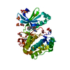
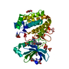
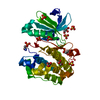
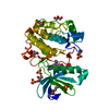
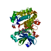
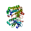
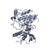
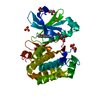
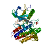
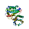
| Entry | Database: PDB / ID: 1uu3 | ||||||
|---|---|---|---|---|---|---|---|
| Title | Structure of human PDK1 kinase domain in complex with LY333531 | ||||||
 Components Components | 3-PHOSPHOINOSITIDE DEPENDENT PROTEIN KINASE-1 | ||||||
 Keywords Keywords | TRANSFERASE / PROTEIN KINASE / PKB / PDK1 / INHIBITOR / LY333531 / DIABETES / CANCER / SERINE/THREONINE-PROTEIN KINASE | ||||||
| Function / homology |  Function and homology information Function and homology informationintracellular signaling cassette / 3-phosphoinositide-dependent protein kinase activity / Activation of AKT2 / regulation of mast cell degranulation / type B pancreatic cell development / negative regulation of toll-like receptor signaling pathway / RSK activation / positive regulation of vascular endothelial cell proliferation / regulation of canonical NF-kappaB signal transduction / phospholipase activator activity ...intracellular signaling cassette / 3-phosphoinositide-dependent protein kinase activity / Activation of AKT2 / regulation of mast cell degranulation / type B pancreatic cell development / negative regulation of toll-like receptor signaling pathway / RSK activation / positive regulation of vascular endothelial cell proliferation / regulation of canonical NF-kappaB signal transduction / phospholipase activator activity / positive regulation of sprouting angiogenesis / Constitutive Signaling by AKT1 E17K in Cancer / negative regulation of cardiac muscle cell apoptotic process / Role of LAT2/NTAL/LAB on calcium mobilization / CD28 dependent PI3K/Akt signaling / negative regulation of endothelial cell apoptotic process / positive regulation of blood vessel endothelial cell migration / SARS-CoV-2 targets host intracellular signalling and regulatory pathways / Estrogen-stimulated signaling through PRKCZ / extrinsic apoptotic signaling pathway / vascular endothelial cell response to laminar fluid shear stress / SARS-CoV-1 targets host intracellular signalling and regulatory pathways / RHO GTPases activate PKNs / GPVI-mediated activation cascade / phospholipase binding / T cell costimulation / Integrin signaling / cell projection / insulin-like growth factor receptor signaling pathway / cellular response to epidermal growth factor stimulus / positive regulation of release of sequestered calcium ion into cytosol / VEGFR2 mediated cell proliferation / VEGFR2 mediated vascular permeability / positive regulation of protein localization to plasma membrane / calcium-mediated signaling / phosphatidylinositol 3-kinase/protein kinase B signal transduction / negative regulation of transforming growth factor beta receptor signaling pathway / CLEC7A (Dectin-1) signaling / epidermal growth factor receptor signaling pathway / FCERI mediated NF-kB activation / cellular response to insulin stimulus / positive regulation of angiogenesis / insulin receptor signaling pathway / Regulation of TP53 Degradation / PIP3 activates AKT signaling / protein autophosphorylation / Downstream TCR signaling / G beta:gamma signalling through PI3Kgamma / cell migration / actin cytoskeleton organization / cytoplasmic vesicle / High laminar flow shear stress activates signaling by PIEZO1 and PECAM1:CDH5:KDR in endothelial cells / protein phosphorylation / positive regulation of phosphatidylinositol 3-kinase/protein kinase B signal transduction / non-specific serine/threonine protein kinase / postsynaptic density / intracellular signal transduction / protein serine kinase activity / focal adhesion / protein serine/threonine kinase activity / DNA-templated transcription / ATP binding / nucleus / plasma membrane / cytosol / cytoplasm Similarity search - Function | ||||||
| Biological species |  HOMO SAPIENS (human) HOMO SAPIENS (human) | ||||||
| Method |  X-RAY DIFFRACTION / SYNCHROTRON / MOLECULAR REPLACEMENT / Resolution: 1.7 Å X-RAY DIFFRACTION / SYNCHROTRON / MOLECULAR REPLACEMENT / Resolution: 1.7 Å | ||||||
 Authors Authors | Komander, D. / Kular, G.S. / Schuttelkopf, A.W. / Deak, M. / Prakash, K.R. / Bain, J. / Elliot, M. / Garrido-Franco, M. / Kozikowski, A.P. / Alessi, D.R. / Van Aalten, D.M.F. | ||||||
 Citation Citation | Journal: Structure / Year: 2004 Title: Interactions of Ly333531 and Other Bisindolyl Maleimide Inhibitors with Pdk1 Authors: Komander, D. / Kular, G.S. / Schuttelkopf, A.W. / Deak, M. / Prakash, K.R. / Bain, J. / Elliot, M. / Garrido-Franco, M. / Kozikowski, A.P. / Alessi, D.R. / Van Aalten, D.M.F. | ||||||
| History |
|
- Structure visualization
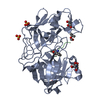
| Structure viewer | Molecule: MolmilJmol/JSmol |
|---|
- Downloads & links
-Download
| PDBx/mmCIF format | 1uu3.cif.gz | 82.8 KB | Display | PDBx/mmCIF format |
|---|---|---|---|---|
| PDB format | pdb1uu3.ent.gz | 61.4 KB | Display | PDB format |
| PDBx/mmJSON format | 1uu3.json.gz | Tree view | PDBx/mmJSON format | |
| Others |  Other downloads Other downloads |
-Validation report
| Arichive directory | https://data.pdbj.org/pub/pdb/validation_reports/uu/1uu3ftp://data.pdbj.org/pub/pdb/validation_reports/uu/1uu3 | HTTPS FTP |
|---|
-Related structure data
| Related structure data | 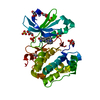 1uu7C 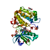 1uu8C 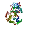 1uu9C  1uvrC 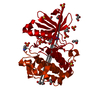 1h1wS S: Starting model for refinement C: citing same article ( |
|---|---|
| Similar structure data |
-Links
 PDBj
PDBj


- Assembly
| Deposited unit | 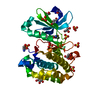
| |||||||||
|---|---|---|---|---|---|---|---|---|---|---|
| 1 |
| |||||||||
| Unit cell |
| |||||||||
| Components on special symmetry positions |
|
-Components
| #1: Protein | Mass: 35498.688 Da / Num. of mol.: 1 / Fragment: KINASE DOMAIN, RESIDUES 51-360 Source method: isolated from a genetically manipulated source Source: (gene. exp.) HOMO SAPIENS (human) / Description: BACULOVIRUS INFECTED / Plasmid: PFASTBAC1 / Cell line (production host): Sf21 / Production host:   SPODOPTERA FRUGIPERDA (fall armyworm) / References: UniProt: O15530, EC: 2.7.1.37 SPODOPTERA FRUGIPERDA (fall armyworm) / References: UniProt: O15530, EC: 2.7.1.37 | ||||||||
|---|---|---|---|---|---|---|---|---|---|
| #2: Chemical | ChemComp-GOL /   Mass: 92.094 Da / Num. of mol.: 8 / Source method: obtained synthetically / Formula: C3H8O3 Mass: 92.094 Da / Num. of mol.: 8 / Source method: obtained synthetically / Formula: C3H8O3#3: Chemical | ChemComp-SO4 /   Mass: 96.063 Da / Num. of mol.: 7 / Source method: obtained synthetically / Formula: SO4 Mass: 96.063 Da / Num. of mol.: 7 / Source method: obtained synthetically / Formula: SO4#4: Chemical | ChemComp-LY4 / ( | 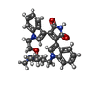  Mass: 468.547 Da / Num. of mol.: 1 / Source method: obtained synthetically / Formula: C28H28N4O3 Mass: 468.547 Da / Num. of mol.: 1 / Source method: obtained synthetically / Formula: C28H28N4O3#5: Water | ChemComp-HOH / |  Mass: 18.015 Da / Num. of mol.: 282 / Source method: isolated from a natural source / Formula: H2O Mass: 18.015 Da / Num. of mol.: 282 / Source method: isolated from a natural source / Formula: H2OHas protein modification | Y | |
-Experimental details
-Experiment
| Experiment | Method: X-RAY DIFFRACTION / Number of used crystals: 1 |
|---|
- Sample preparation
| Crystal | Density Matthews: 3 Å3/Da / Density % sol: 58 % | ||||||||||||||||||||
|---|---|---|---|---|---|---|---|---|---|---|---|---|---|---|---|---|---|---|---|---|---|
| Crystal grow | pH: 7.5 / Details: 2.1 M AMMONIUM SULPHATE 0.1 M TRIS-HCL PH 7.2 | ||||||||||||||||||||
| Crystal grow | *PLUS Method: vapor diffusion, sitting drop | ||||||||||||||||||||
| Components of the solutions | *PLUS
|
-Data collection
| Diffraction | Mean temperature: 100 K |
|---|---|
| Diffraction source | Source: SYNCHROTRON / Site: ESRF  / Beamline: ID14-2 / Wavelength: 0.933 / Beamline: ID14-2 / Wavelength: 0.933 |
| Detector | Type: ADSC CCD / Detector: CCD / Date: Feb 15, 2003 |
| Radiation | Protocol: SINGLE WAVELENGTH / Monochromatic (M) / Laue (L): M / Scattering type: x-ray |
| Radiation wavelength | Wavelength: 0.933 Å / Relative weight: 1 |
| Reflection | Resolution: 1.7→25 Å / Num. obs: 44644 / % possible obs: 99.9 % / Redundancy: 4.9 % / Rmerge(I) obs: 0.044 / Net I/σ(I): 22.1 |
| Reflection shell | Resolution: 1.7→1.76 Å / Redundancy: 3.7 % / Rmerge(I) obs: 0.278 / Mean I/σ(I) obs: 4.7 / % possible all: 98.6 |
| Reflection | *PLUS Highest resolution: 1.7 Å / Lowest resolution: 25 Å / Redundancy: 4.9 % / Num. measured all: 218856 / Rmerge(I) obs: 0.044 |
| Reflection shell | *PLUS % possible obs: 98.6 % / Redundancy: 3.7 % / Num. unique obs: 4344 / Num. measured obs: 15901 / Rmerge(I) obs: 0.278 / Mean I/σ(I) obs: 4.7 |
- Processing
| Software |
| ||||||||||||||||||||||||||||||||||||||||||||||||||||||||||||||||||||||||||||||||||||||||||||||||||||||||||||||||||||||||||||||||||||||||||||||||||||||||||||||||||||||||||||||||||||||
|---|---|---|---|---|---|---|---|---|---|---|---|---|---|---|---|---|---|---|---|---|---|---|---|---|---|---|---|---|---|---|---|---|---|---|---|---|---|---|---|---|---|---|---|---|---|---|---|---|---|---|---|---|---|---|---|---|---|---|---|---|---|---|---|---|---|---|---|---|---|---|---|---|---|---|---|---|---|---|---|---|---|---|---|---|---|---|---|---|---|---|---|---|---|---|---|---|---|---|---|---|---|---|---|---|---|---|---|---|---|---|---|---|---|---|---|---|---|---|---|---|---|---|---|---|---|---|---|---|---|---|---|---|---|---|---|---|---|---|---|---|---|---|---|---|---|---|---|---|---|---|---|---|---|---|---|---|---|---|---|---|---|---|---|---|---|---|---|---|---|---|---|---|---|---|---|---|---|---|---|---|---|---|---|
| Refinement | Method to determine structure: MOLECULAR REPLACEMENT Starting model: PDB ENTRY 1H1W Resolution: 1.7→25 Å / Cor.coef. Fo:Fc: 0.947 / Cor.coef. Fo:Fc free: 0.937 / SU B: 1.428 / SU ML: 0.049 / Cross valid method: THROUGHOUT / ESU R: 0.087 / ESU R Free: 0.088 / Stereochemistry target values: MAXIMUM LIKELIHOOD Details: HYDROGENS HAVE BEEN ADDED IN THE RIDING POSITIONS SOME SIDECHAINS REFINED WITH OCCUPANCY = 0.00 DUE TO DISORDER. SOME RESIDUES MUTATED TO ALANINE DUE TO DISORDER.
| ||||||||||||||||||||||||||||||||||||||||||||||||||||||||||||||||||||||||||||||||||||||||||||||||||||||||||||||||||||||||||||||||||||||||||||||||||||||||||||||||||||||||||||||||||||||
| Solvent computation | Ion probe radii: 0.8 Å / Shrinkage radii: 0.8 Å / VDW probe radii: 1.4 Å / Solvent model: BABINET MODEL WITH MASK | ||||||||||||||||||||||||||||||||||||||||||||||||||||||||||||||||||||||||||||||||||||||||||||||||||||||||||||||||||||||||||||||||||||||||||||||||||||||||||||||||||||||||||||||||||||||
| Displacement parameters | Biso mean: 17.71 Å2
| ||||||||||||||||||||||||||||||||||||||||||||||||||||||||||||||||||||||||||||||||||||||||||||||||||||||||||||||||||||||||||||||||||||||||||||||||||||||||||||||||||||||||||||||||||||||
| Refinement step | Cycle: LAST / Resolution: 1.7→25 Å
| ||||||||||||||||||||||||||||||||||||||||||||||||||||||||||||||||||||||||||||||||||||||||||||||||||||||||||||||||||||||||||||||||||||||||||||||||||||||||||||||||||||||||||||||||||||||
| Refine LS restraints |
|