[English] 日本語
Yorodumi
- PDB-1oex: Atomic Resolution Structure of Endothiapepsin in Complex with a H... -

+
Open data


ID or keywords:

Loading...

-
Basic information

Entry
Database: PDB / ID: 1oex
TitleAtomic Resolution Structure of Endothiapepsin in Complex with a Hydroxyethylene Transition State Analogue Inhibitor H261
Components
  • ENDOTHIAPEPSIN
  • INHIBITOR H261
KeywordsHYDROLASE/HYDROLASE INHIBITOR / ASPARTIC PROTEINASE MECHANISM / ATOMIC RESOLUTION / SUCCINIMIDE / ANISOTROPIC REFINEMENT / HYDROLASE-HYDROLASE INHIBITOR COMPLEX
Function / homology
Function and homology information


endothiapepsin / aspartic-type endopeptidase activity / proteolysis
Similarity search - Function
Aspergillopepsin-like catalytic domain / Eukaryotic aspartyl protease / Aspartic peptidase A1 family / Peptidase family A1 domain / Peptidase family A1 domain profile. / Cathepsin D, subunit A; domain 1 / Acid Proteases / Aspartic peptidase, active site / Eukaryotic and viral aspartyl proteases active site. / Aspartic peptidase domain superfamily ...Aspergillopepsin-like catalytic domain / Eukaryotic aspartyl protease / Aspartic peptidase A1 family / Peptidase family A1 domain / Peptidase family A1 domain profile. / Cathepsin D, subunit A; domain 1 / Acid Proteases / Aspartic peptidase, active site / Eukaryotic and viral aspartyl proteases active site. / Aspartic peptidase domain superfamily / Beta Barrel / Mainly Beta
Similarity search - Domain/homology
H261 INHIBITOR / Endothiapepsin
Similarity search - Component
Biological speciesCRYPHONECTRIA PARASITICA (chestnut blight fungus)
SYNTHETIC CONSTRUCT (others)
MethodX-RAY DIFFRACTION / SYNCHROTRON / OTHER / Resolution: 1.1 Å
AuthorsCoates, L. / Erskine, P.T. / Mall, S. / Gill, R.S. / Wood, S.P. / Myles, D.A.A. / Cooper, J.B.
CitationJournal: Protein Sci. / Year: 2003
Title: Atomic Resolution Analysis of the Catalytic Site of an Aspartic Proteinase and an Unexpected Mode of Binding by Short Peptides
Authors: Erskine, P.T. / Coates, L. / Mall, S. / Gill, R.S. / Wood, S.P. / Myles, D.A.A. / Cooper, J.B.
History
DepositionMar 31, 2003Deposition site: PDBE / Processing site: PDBE
Revision 1.0Apr 2, 2003Provider: repository / Type: Initial release
Revision 1.1Jul 13, 2011Group: Atomic model / Database references ...Atomic model / Database references / Derived calculations / Non-polymer description / Structure summary / Version format compliance
Revision 1.2Nov 30, 2012Group: Other
Revision 1.3Feb 8, 2017Group: Source and taxonomy
Revision 1.4Jul 24, 2019Group: Data collection / Derived calculations / Category: diffrn_source / struct_conn
Item: _diffrn_source.pdbx_synchrotron_site / _struct_conn.pdbx_leaving_atom_flag
Revision 2.0Nov 15, 2023Group: Atomic model / Data collection ...Atomic model / Data collection / Database references / Derived calculations / Other
Category: atom_site / atom_site_anisotrop ...atom_site / atom_site_anisotrop / chem_comp_atom / chem_comp_bond / database_2 / pdbx_database_status / pdbx_validate_main_chain_plane / pdbx_validate_peptide_omega / pdbx_validate_rmsd_angle / struct_conn / struct_site
Item: _atom_site.auth_atom_id / _atom_site.label_atom_id ..._atom_site.auth_atom_id / _atom_site.label_atom_id / _atom_site_anisotrop.pdbx_auth_atom_id / _atom_site_anisotrop.pdbx_label_atom_id / _database_2.pdbx_DOI / _database_2.pdbx_database_accession / _pdbx_database_status.status_code_sf / _pdbx_validate_peptide_omega.auth_comp_id_1 / _pdbx_validate_peptide_omega.auth_comp_id_2 / _pdbx_validate_peptide_omega.auth_seq_id_1 / _pdbx_validate_peptide_omega.auth_seq_id_2 / _pdbx_validate_peptide_omega.omega / _pdbx_validate_rmsd_angle.angle_deviation / _pdbx_validate_rmsd_angle.angle_standard_deviation / _pdbx_validate_rmsd_angle.angle_target_value / _pdbx_validate_rmsd_angle.angle_value / _pdbx_validate_rmsd_angle.auth_asym_id_1 / _pdbx_validate_rmsd_angle.auth_asym_id_2 / _pdbx_validate_rmsd_angle.auth_asym_id_3 / _pdbx_validate_rmsd_angle.auth_atom_id_1 / _pdbx_validate_rmsd_angle.auth_atom_id_2 / _pdbx_validate_rmsd_angle.auth_atom_id_3 / _pdbx_validate_rmsd_angle.auth_comp_id_1 / _pdbx_validate_rmsd_angle.auth_comp_id_2 / _pdbx_validate_rmsd_angle.auth_comp_id_3 / _pdbx_validate_rmsd_angle.auth_seq_id_1 / _pdbx_validate_rmsd_angle.auth_seq_id_2 / _pdbx_validate_rmsd_angle.auth_seq_id_3 / _pdbx_validate_rmsd_angle.label_alt_id_1 / _pdbx_validate_rmsd_angle.label_alt_id_2 / _pdbx_validate_rmsd_angle.label_alt_id_3 / _pdbx_validate_rmsd_angle.linker_flag / _struct_conn.pdbx_leaving_atom_flag / _struct_site.pdbx_auth_asym_id / _struct_site.pdbx_auth_comp_id / _struct_site.pdbx_auth_seq_id
Remark 700 
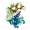
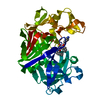
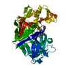
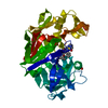
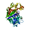
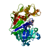
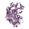
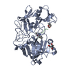
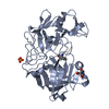
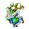
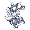
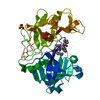
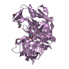
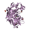
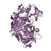
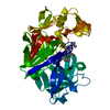
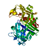
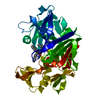
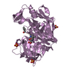
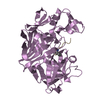
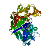
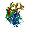
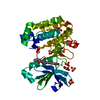
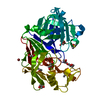
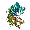
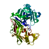
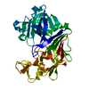
SHEET THE SHEET STRUCTURE OF THIS MOLECULE IS BIFURCATED. IN ORDER TO REPRESENT THIS FEATURE IN ... SHEET THE SHEET STRUCTURE OF THIS MOLECULE IS BIFURCATED. IN ORDER TO REPRESENT THIS FEATURE IN THE SHEET RECORDS BELOW, TWO SHEETS ARE DEFINED.

-
Structure visualization

Structure viewerMolecule:
MolmilJmol/JSmol

Downloads & links

-
Assembly

Deposited unit
A: ENDOTHIAPEPSIN
B: INHIBITOR H261
hetero molecules


Theoretical massNumber of molelcules
Total (without water)35,3087
Polymers34,8322
Non-polymers4765
Water8,989499
1


  • Idetical with deposited unit
  • defined by author&software
TypeNameSymmetry operationNumber
identity operation1_555x,y,z1
Buried area2610 Å2
ΔGint-51 kcal/mol
Surface area12920 Å2
MethodPISA
Unit cell
Length a, b, c (Å)42.580, 74.510, 42.350
Angle α, β, γ (deg.)90.00, 97.56, 90.00
Int Tables number4
Space group name H-MP1211

-
Components

#1: Protein ENDOTHIAPEPSIN / ASPARTATE PROTEASE / EAPA / EPN-1


Mass: 33795.840 Da / Num. of mol.: 1 / Fragment: RESIDUES 90-419 / Source method: isolated from a natural source
Details: HYDROXYETHYLENE TRANSITION STATE ANALOGUE INHIBITOR H261 BOUND AT ACTIVE SITE
Source: (natural) CRYPHONECTRIA PARASITICA (chestnut blight fungus)
References: UniProt: P11838, endothiapepsin
#2: Protein/peptide INHIBITOR H261


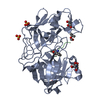
Type: Peptide-like / Class: Inhibitor / Mass: 1036.266 Da / Num. of mol.: 1 / Source method: obtained synthetically / Details: BOC-HIS-PRO-PHE-ALA-LOV-ILE-HIS / Source: (synth.) SYNTHETIC CONSTRUCT (others) / References: H261 INHIBITOR
#3: Chemical ChemComp-GOL / GLYCEROL / GLYCERIN / PROPANE-1,2,3-TRIOL


Mass: 92.094 Da / Num. of mol.: 1 / Source method: obtained synthetically / Formula: C3H8O3
#4: Chemical
ChemComp-SO4 / SULFATE ION


Mass: 96.063 Da / Num. of mol.: 4 / Source method: obtained synthetically / Formula: SO4
#5: Water ChemComp-HOH / water


Mass: 18.015 Da / Num. of mol.: 499 / Source method: isolated from a natural source / Formula: H2O
Sequence detailsASP A54 AND GLY A55 HAVE CYCLISED TO FORM A SUCCINIMIDE

-
Experimental details

-
Experiment

ExperimentMethod: X-RAY DIFFRACTION / Number of used crystals: 1

-
Sample preparation

CrystalDensity Matthews: 1.99 Å3/Da / Density % sol: 38 %
Crystal growpH: 4.6
Details: BATCH METHOD WITH ENZYME AT A CONCENTRATION OF 2 MG/ML IN 100 MM SODIUM ACETATE BUFFER AT PH 4.6 (MOEWS AND BUNN, 1970). CRYSTALS LEFT TO GROW FOLLOWING SLOW ADDITION OF AMMONIUM SULPHATE TO ...Details: BATCH METHOD WITH ENZYME AT A CONCENTRATION OF 2 MG/ML IN 100 MM SODIUM ACETATE BUFFER AT PH 4.6 (MOEWS AND BUNN, 1970). CRYSTALS LEFT TO GROW FOLLOWING SLOW ADDITION OF AMMONIUM SULPHATE TO FINAL CONCENTRATION OF 0.35 G/ML (55% SATURATION).

-
Data collection

DiffractionMean temperature: 100 K
Diffraction sourceSource: SYNCHROTRON / Site: EMBL/DESY, HAMBURG / Beamline: BW7B / Wavelength: 0.834
DetectorType: MARRESEARCH / Detector: IMAGE PLATE / Date: Jun 15, 1998
RadiationProtocol: SINGLE WAVELENGTH / Monochromatic (M) / Laue (L): M / Scattering type: x-ray
Radiation wavelengthWavelength: 0.834 Å / Relative weight: 1
ReflectionResolution: 1.1→15.6 Å / Num. obs: 105807 / % possible obs: 99.8 % / Redundancy: 3.7 % / Rmerge(I) obs: 0.125 / Net I/σ(I): 9.8
Reflection shellResolution: 1.1→1.14 Å / Redundancy: 3.5 % / Rmerge(I) obs: 0.32 / Mean I/σ(I) obs: 4.8 / % possible all: 99.5

-
Processing

Software
NameClassification
SHELXL-97refinement
MOSFLMdata reduction
Agrovatadata scaling
ROTAVATAdata scaling
SHELXL-97phasing
RefinementMethod to determine structure: OTHER / Resolution: 1.1→100 Å / Num. parameters: 27154 / Num. restraintsaints: 33073 / Cross valid method: THROUGHOUT / σ(F): 0 / Stereochemistry target values: ENGH AND HUBER
RfactorNum. reflection% reflectionSelection details
Rfree0.178 5285 5 %RANDOM
obs0.14 -99.8 %-
all-105807 --
Solvent computationSolvent model: MOEWS AND KRETSINGER
Refine analyzeNum. disordered residues: 25
Refinement stepCycle: LAST / Resolution: 1.1→100 Å
ProteinNucleic acidLigandSolventTotal
Num. atoms2462 0 26 499 2987
Refine LS restraints
Refine-IDTypeDev ideal
X-RAY DIFFRACTIONs_bond_d0.013
X-RAY DIFFRACTIONs_angle_d0.028
X-RAY DIFFRACTIONs_similar_dist0
X-RAY DIFFRACTIONs_from_restr_planes0.032
X-RAY DIFFRACTIONs_zero_chiral_vol0.079
X-RAY DIFFRACTIONs_non_zero_chiral_vol0.078
X-RAY DIFFRACTIONs_anti_bump_dis_restr0.074
X-RAY DIFFRACTIONs_rigid_bond_adp_cmpnt0.005
X-RAY DIFFRACTIONs_similar_adp_cmpnt0.03
X-RAY DIFFRACTIONs_approx_iso_adps0.071

+
About Yorodumi

-
News

-
Feb 9, 2022. New format data for meta-information of EMDB entries

New format data for meta-information of EMDB entries

  • Version 3 of the EMDB header file is now the official format.
  • The previous official version 1.9 will be removed from the archive.

Related info.:EMDB header

External links:wwPDB to switch to version 3 of the EMDB data model

-
Aug 12, 2020. Covid-19 info

Covid-19 info

URL: https://pdbjlvh1.pdbj.org/emnavi/covid19.php

New page: Covid-19 featured information page in EM Navigator.

Related info.:Covid-19 info / Mar 5, 2020. Novel coronavirus structure data

+
Mar 5, 2020. Novel coronavirus structure data

Novel coronavirus structure data

Related info.:Yorodumi Speices / Aug 12, 2020. Covid-19 info

External links:COVID-19 featured content - PDBj / Molecule of the Month (242):Coronavirus Proteases

+
Jan 31, 2019. EMDB accession codes are about to change! (news from PDBe EMDB page)

EMDB accession codes are about to change! (news from PDBe EMDB page)

  • The allocation of 4 digits for EMDB accession codes will soon come to an end. Whilst these codes will remain in use, new EMDB accession codes will include an additional digit and will expand incrementally as the available range of codes is exhausted. The current 4-digit format prefixed with “EMD-” (i.e. EMD-XXXX) will advance to a 5-digit format (i.e. EMD-XXXXX), and so on. It is currently estimated that the 4-digit codes will be depleted around Spring 2019, at which point the 5-digit format will come into force.
  • The EM Navigator/Yorodumi systems omit the EMD- prefix.

Related info.:Q: What is EMD? / ID/Accession-code notation in Yorodumi/EM Navigator

External links:EMDB Accession Codes are Changing Soon! / Contact to PDBj

+
Jul 12, 2017. Major update of PDB

Major update of PDB

  • wwPDB released updated PDB data conforming to the new PDBx/mmCIF dictionary.
  • This is a major update changing the version number from 4 to 5, and with Remediation, in which all the entries are updated.
  • In this update, many items about electron microscopy experimental information are reorganized (e.g. em_software).
  • Now, EM Navigator and Yorodumi are based on the updated data.

External links:wwPDB Remediation / Enriched Model Files Conforming to OneDep Data Standards Now Available in the PDB FTP Archive

-
Yorodumi

Thousand views of thousand structures

  • Yorodumi is a browser for structure data from EMDB, PDB, SASBDB, etc.
  • This page is also the successor to EM Navigator detail page, and also detail information page/front-end page for Omokage search.
  • The word "yorodu" (or yorozu) is an old Japanese word meaning "ten thousand". "mi" (miru) is to see.

Related info.:EMDB / PDB / SASBDB / Comparison of 3 databanks / Yorodumi Search / Aug 31, 2016. New EM Navigator & Yorodumi / Yorodumi Papers / Jmol/JSmol / Function and homology information / Changes in new EM Navigator and Yorodumi

Read more