 Movie
Movie Controller


+ Open data

- Basic information
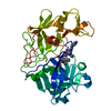
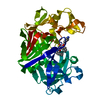
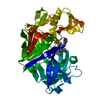
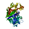
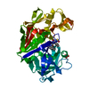
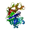
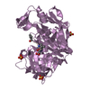
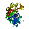
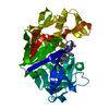
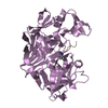
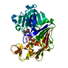
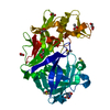
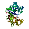
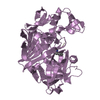
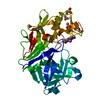
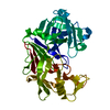
| Entry | Database: PDB / ID: 1gvx | |||||||||
|---|---|---|---|---|---|---|---|---|---|---|
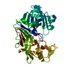
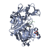
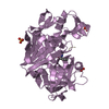
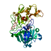
| Title | Endothiapepsin complexed with H256 | |||||||||
 Components Components |
| |||||||||
 Keywords Keywords | HYDROLASE/HYDROLASE INHIBITOR / HYDROLASE / ASPARTIC PROTEINASE MECHANISM / TETRAHEDRAL INTERMEDIATE / HYDROLASE-HYDROLASE INHIBITOR COMPLEX | |||||||||
| Function / homology |  Function and homology information Function and homology information | |||||||||
| Biological species |  ENDOTHIA PARASITICA (chestnut blight fungus) ENDOTHIA PARASITICA (chestnut blight fungus)SYNTHETIC CONSTRUCT (others) | |||||||||
| Method |  X-RAY DIFFRACTION / SYNCHROTRON / MOLECULAR REPLACEMENT / Resolution: 1 Å X-RAY DIFFRACTION / SYNCHROTRON / MOLECULAR REPLACEMENT / Resolution: 1 Å | |||||||||
 Authors Authors | Coates, L. / Erskine, P.T. / Crump, M.P. / Wood, S.P. / Cooper, J.B. | |||||||||
 Citation Citation | Journal: J.Mol.Biol. / Year: 2002 Title: Five Atomic Resolution Structures of Endothiapepsin Inhibitor Complexes: Implications for the Aspartic Proteinase Mechanism Authors: Coates, L. / Erskine, P.T. / Crump, M.P. / Wood, S.P. / Cooper, J.B. | |||||||||
| History |
| |||||||||
| Remark 700 | SHEET THE SHEET STRUCTURE OF THIS MOLECULE IS BIFURCATED. IN ORDER TO REPRESENT THIS FEATURE IN ... SHEET THE SHEET STRUCTURE OF THIS MOLECULE IS BIFURCATED. IN ORDER TO REPRESENT THIS FEATURE IN THE SHEET RECORDS BELOW, TWO SHEETS ARE DEFINED. |
- Structure visualization
| Structure viewer | Molecule: MolmilJmol/JSmol |
|---|
- Downloads & links
-Download
| PDBx/mmCIF format | 1gvx.cif.gz | 152.5 KB | Display | PDBx/mmCIF format |
|---|---|---|---|---|
| PDB format | pdb1gvx.ent.gz | 119.1 KB | Display | PDB format |
| PDBx/mmJSON format | 1gvx.json.gz | Tree view | PDBx/mmJSON format | |
| Others |  Other downloads Other downloads |
-Validation report
| Arichive directory | https://data.pdbj.org/pub/pdb/validation_reports/gv/1gvxftp://data.pdbj.org/pub/pdb/validation_reports/gv/1gvx | HTTPS FTP |
|---|
-Related structure data
| Related structure data |  1gvtC 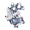 1gvuC 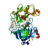 1gvvC  1gvwC  3er5S C: citing same article ( S: Starting model for refinement |
|---|---|
| Similar structure data |


-Links
 PDBj
PDBj
- Assembly
| Deposited unit | 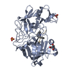
| ||||||||
|---|---|---|---|---|---|---|---|---|---|
| 1 |
| ||||||||
| Unit cell |
|
-Components
| #1: Protein | Mass: 33795.840 Da / Num. of mol.: 1 / Source method: isolated from a natural source Source: (natural) ENDOTHIA PARASITICA (chestnut blight fungus)References: UniProt: P11838, endothiapepsin | ||
|---|---|---|---|
| #2: Protein/peptide |   Type: Peptide-like / Class: Inhibitor / Mass: 912.019 Da / Num. of mol.: 1 / Source method: obtained synthetically / Source: (synth.) SYNTHETIC CONSTRUCT (others) Type: Peptide-like / Class: Inhibitor / Mass: 912.019 Da / Num. of mol.: 1 / Source method: obtained synthetically / Source: (synth.) SYNTHETIC CONSTRUCT (others)References: N-{(2R)-3-phenyl-2-[(L-prolyl-L-threonyl-L-alpha-glutamyl)amino]propyl}-L-phenylalanyl-N~5~-[amino(iminio)methyl]- L-ornithyl-L-glutamic acid | ||
| #3: Chemical |   Mass: 96.063 Da / Num. of mol.: 2 / Source method: obtained synthetically / Formula: SO4 Mass: 96.063 Da / Num. of mol.: 2 / Source method: obtained synthetically / Formula: SO4#4: Water | ChemComp-HOH / |  Mass: 18.015 Da / Num. of mol.: 399 / Source method: isolated from a natural source / Formula: H2O Mass: 18.015 Da / Num. of mol.: 399 / Source method: isolated from a natural source / Formula: H2O |
-Experimental details
-Experiment
| Experiment | Method: X-RAY DIFFRACTION / Number of used crystals: 1 |
|---|
- Sample preparation
| Crystal | Density Matthews: 2.04 Å3/Da / Density % sol: 39.83 % | |||||||||||||||
|---|---|---|---|---|---|---|---|---|---|---|---|---|---|---|---|---|
| Crystal grow | pH: 4.5 / Details: PH 4.50 | |||||||||||||||
| Crystal grow | *PLUS Method: unknown | |||||||||||||||
| Components of the solutions | *PLUS
|
-Data collection
| Diffraction | Mean temperature: 130 K |
|---|---|
| Diffraction source | Source: SYNCHROTRON / Site: ESRF  / Beamline: ID14-2 / Wavelength: 0.933 / Beamline: ID14-2 / Wavelength: 0.933 |
| Detector | Type: ADSC CCD / Detector: CCD / Date: Apr 1, 2001 |
| Radiation | Protocol: SINGLE WAVELENGTH / Monochromatic (M) / Laue (L): M / Scattering type: x-ray |
| Radiation wavelength | Wavelength: 0.933 Å / Relative weight: 1 |
| Reflection | Resolution: 1→10 Å / Num. obs: 136765 / % possible obs: 96.9 % / Redundancy: 6.9 % / Rmerge(I) obs: 0.079 / Net I/σ(I): 12.7 |
| Reflection shell | Resolution: 1→1.05 Å / Redundancy: 6.1 % / Rmerge(I) obs: 0.163 / Mean I/σ(I) obs: 3.6 / % possible all: 94.3 |
| Reflection | *PLUS Lowest resolution: 10 Å |
| Reflection shell | *PLUS % possible obs: 94.3 % |
- Processing
| Software |
| |||||||||||||||||||||||||||||||||
|---|---|---|---|---|---|---|---|---|---|---|---|---|---|---|---|---|---|---|---|---|---|---|---|---|---|---|---|---|---|---|---|---|---|---|
| Refinement | Method to determine structure: MOLECULAR REPLACEMENT Starting model: PDB ENTRY 3ER5 Resolution: 1→10 Å / Cross valid method: THROUGHOUT / σ(F): 0 / Stereochemistry target values: ENGH AND HUBER
| |||||||||||||||||||||||||||||||||
| Solvent computation | Solvent model: MOEWS AND KRETSINGER | |||||||||||||||||||||||||||||||||
| Refinement step | Cycle: LAST / Resolution: 1→10 Å
| |||||||||||||||||||||||||||||||||
| Refine LS restraints |
| |||||||||||||||||||||||||||||||||
| Refinement | *PLUS Lowest resolution: 10 Å / Rfactor Rfree: 0.1647 / Rfactor Rwork: 0.1393 | |||||||||||||||||||||||||||||||||
| Solvent computation | *PLUS | |||||||||||||||||||||||||||||||||
| Displacement parameters | *PLUS |
