 Movie
Movie Controller

[English] 日本語
 Yorodumi
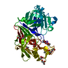
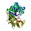
- PDB-4er4: HIGH-RESOLUTION X-RAY ANALYSES OF RENIN INHIBITOR-ASPARTIC PROTEI... -
+ Open data

- Basic information
| Entry | Database: PDB / ID: 4er4 | |||||||||
|---|---|---|---|---|---|---|---|---|---|---|
| Title | HIGH-RESOLUTION X-RAY ANALYSES OF RENIN INHIBITOR-ASPARTIC PROTEINASE COMPLEXES | |||||||||
 Components Components |
| |||||||||
 Keywords Keywords | HYDROLASE/HYDROLASE INHIBITOR / HYDROLASE / ACID PROTEINASE / HYDROLASE-HYDROLASE INHIBITOR COMPLEX | |||||||||
| Function / homology |  Function and homology information Function and homology information | |||||||||
| Biological species |  Cryphonectria parasitica (chestnut blight fungus) Cryphonectria parasitica (chestnut blight fungus) | |||||||||
| Method |  X-RAY DIFFRACTION / Resolution: 2.1 Å X-RAY DIFFRACTION / Resolution: 2.1 Å | |||||||||
 Authors Authors | Foundling, S.I. / Watson, F.E. / Szelke, M. / Blundell, T.L. | |||||||||
 Citation Citation | Journal: Nature / Year: 1987 Title: High resolution X-ray analyses of renin inhibitor-aspartic proteinase complexes. Authors: Foundling, S.I. / Cooper, J. / Watson, F.E. / Cleasby, A. / Pearl, L.H. / Sibanda, B.L. / Hemmings, A. / Wood, S.P. / Blundell, T.L. / Valler, M.J. / Norey, C.G. / Kay, J. / Boger, J. / ...Authors: Foundling, S.I. / Cooper, J. / Watson, F.E. / Cleasby, A. / Pearl, L.H. / Sibanda, B.L. / Hemmings, A. / Wood, S.P. / Blundell, T.L. / Valler, M.J. / Norey, C.G. / Kay, J. / Boger, J. / Dunn, B.M. / Leckieparallel, B.J. / Jone, D.M. / Atrash, B. / Hallett, A. / Szelke, M. #1: Journal: FEBS Lett. / Year: 1984Title: The Active Site of Aspartic Proteinases Authors: Pearl, L. / Blundell, T. #2: Journal: Proc.FEBS Meet. / Year: 1979Title: Active Site of Acid Proteinases Authors: Blundell, T.L. / Jones, H.B. / Khan, G. / Taylor, G. / Sewell, T.S. / Pearl, L.H. / Wood, S.P. #3: Journal: Proc.FEBS Meet. / Year: 1979Title: The Three-Dimensional Structure of Acid Proteinases Authors: Blundell, T.L. / Jenkins, J.A. / Khan, G. / Roychowdhury, P. / Sewell, T. / Tickle, I.J. / Wood, E.A. #4: Journal: Biochim.Biophys.Acta / Year: 1979Title: Four-Fold Structural Repeat in the Acid Proteases Authors: Blundell, T.L. / Sewell, B.T. / Mclachlan, A.D. #5: Journal: Nature / Year: 1978Title: Structural Evidence for Gene Duplication in the Evolution of Acid Proteases Authors: Tang, J. / James, M.N.G. / Hsu, I.N. / Jenkins, J.A. / Blundell, T.L. #6: Journal: Proc.Natl.Acad.Sci.USA / Year: 1977Title: Homology Among Acid Proteases. Comparison of Crystal Structures at 3 Angstroms Resolution of Acid Proteases from Rhizopus Chinensis and Endothia Parasitica Authors: Subramanian, E. / Swan, I.D.A. / Liu, M. / Davies, D.R. / Jenkins, J.A. / Tickle, I.J. / Blundell, T.L. #7: Journal: Adv.Exp.Med.Biol. / Year: 1977Title: X-Ray Analysis and Circular Dichroism of the Acid Protease from Endothia Parasitica and Chymosin Authors: Jenkins, J. / Tickle, I. / Sewell, T. / Ungaretti, L. / Wollmer, A. / Blundell, T. | |||||||||
| History |
|
- Structure visualization
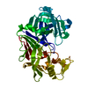
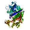
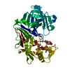
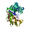
| Structure viewer | Molecule: MolmilJmol/JSmol |
|---|
- Downloads & links
-Download
| PDBx/mmCIF format | 4er4.cif.gz | 81.4 KB | Display | PDBx/mmCIF format |
|---|---|---|---|---|
| PDB format | pdb4er4.ent.gz | 58.7 KB | Display | PDB format |
| PDBx/mmJSON format | 4er4.json.gz | Tree view | PDBx/mmJSON format | |
| Others |  Other downloads Other downloads |
-Validation report
| Arichive directory | https://data.pdbj.org/pub/pdb/validation_reports/er/4er4ftp://data.pdbj.org/pub/pdb/validation_reports/er/4er4 | HTTPS FTP |
|---|
-Related structure data
| Similar structure data |
|---|


-Links
 PDBj
PDBj
- Assembly
| Deposited unit | 
| ||||||||
|---|---|---|---|---|---|---|---|---|---|
| 1 |
| ||||||||
| Unit cell |
| ||||||||
| Atom site foot note | 1: RESIDUES PRO E 23 AND PRO E 133 ARE CIS PROLINES. 2: THE PEPTIDE BOND BETWEEN LEU I 6 AND VAL I 7 HAS BEEN REDUC TO PRODUCE THE SEQUENCE LEU I 6 PSI(CH2-NH) VAL I 7 |
-Components
| #1: Protein | Mass: 33813.855 Da / Num. of mol.: 1 Source method: isolated from a genetically manipulated source Source: (gene. exp.) Cryphonectria parasitica (chestnut blight fungus)References: UniProt: P11838, EC: 3.4.23.6 |
|---|---|
| #2: Protein/peptide | Mass: 1214.501 Da / Num. of mol.: 1 / Source method: obtained synthetically |
| #3: Water | ChemComp-HOH /  Mass: 18.015 Da / Num. of mol.: 325 / Source method: isolated from a natural source / Formula: H2O Mass: 18.015 Da / Num. of mol.: 325 / Source method: isolated from a natural source / Formula: H2O |
| Compound details | THE PEPTIDE BOND BETWEEN RESIDUE LEU I 6 AND RESIDUE VAL I 7 HAS BEEN REDUCED TO CH2-NH2. IT IS ...THE PEPTIDE BOND BETWEEN RESIDUE LEU I 6 AND RESIDUE VAL I 7 HAS BEEN REDUCED TO CH2-NH2. IT IS REPRESENTE |
| Has protein modification | Y |
| Sequence details | THE COMPLETE SEQUENCE WAS DETERMINED BY V. PEDERSEN AS TRYPTIC FRAGMENTS WHICH WERE ALIGNED IN THE ...THE COMPLETE SEQUENCE WAS DETERMINED |
-Experimental details
-Experiment
| Experiment | Method: X-RAY DIFFRACTION |
|---|
- Sample preparation
| Crystal | Density Matthews: 2.43 Å3/Da / Density % sol: 49.46 % | ||||||||||||||||||||||||
|---|---|---|---|---|---|---|---|---|---|---|---|---|---|---|---|---|---|---|---|---|---|---|---|---|---|
| Crystal grow | *PLUS pH: 6.3 / Method: unknown | ||||||||||||||||||||||||
| Components of the solutions | *PLUS
|
-Data collection
| Radiation | Scattering type: x-ray |
|---|---|
| Radiation wavelength | Relative weight: 1 |
- Processing
| Software | Name: PROLSQ / Classification: refinement | ||||||||||||||||||||||||||||||||||||||||||||||||||||||||||||
|---|---|---|---|---|---|---|---|---|---|---|---|---|---|---|---|---|---|---|---|---|---|---|---|---|---|---|---|---|---|---|---|---|---|---|---|---|---|---|---|---|---|---|---|---|---|---|---|---|---|---|---|---|---|---|---|---|---|---|---|---|---|
| Refinement | Resolution: 2.1→20 Å / Rfactor Rwork: 0.194 Details: THE QUANTITY GIVEN IN THE TEMPERATURE FACTOR FIELD OF THE *ATOM* AND *HETATM* RECORDS BELOW IS U**2, WHICH IS THE MEAN-SQUARE AMPLITUDE OF ATOMIC VIBRATION. THE TEMPERATURE FACTOR, B, CAN BE ...Details: THE QUANTITY GIVEN IN THE TEMPERATURE FACTOR FIELD OF THE *ATOM* AND *HETATM* RECORDS BELOW IS U**2, WHICH IS THE MEAN-SQUARE AMPLITUDE OF ATOMIC VIBRATION. THE TEMPERATURE FACTOR, B, CAN BE DERIVED BY THE FOLLOWING RELATION - B = 8 * (PI)**2 * U**2. IT IS AN INDICATION OF POSSIBLE ERRORS IN THE REFINEMENT THAT SOME ARE SLIGHTLY NEGATIVE. | ||||||||||||||||||||||||||||||||||||||||||||||||||||||||||||
| Refinement step | Cycle: LAST / Resolution: 2.1→20 Å
| ||||||||||||||||||||||||||||||||||||||||||||||||||||||||||||
| Refine LS restraints |
| ||||||||||||||||||||||||||||||||||||||||||||||||||||||||||||
| Refinement | *PLUS Rfactor obs: 0.194 | ||||||||||||||||||||||||||||||||||||||||||||||||||||||||||||
| Solvent computation | *PLUS | ||||||||||||||||||||||||||||||||||||||||||||||||||||||||||||
| Displacement parameters | *PLUS | ||||||||||||||||||||||||||||||||||||||||||||||||||||||||||||
| Refine LS restraints | *PLUS Type: o_plane_restr / Dev ideal: 0.013 |
