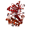
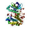
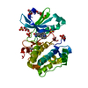
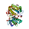
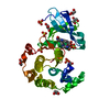
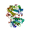
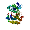
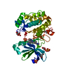
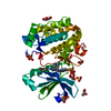
Entry Database : PDB / ID : 1okyTitle Structure of human PDK1 kinase domain in complex with staurosporine 3-PHOSPHOINOSITIDE DEPENDENT PROTEIN KINASE 1 Keywords / / / / Function / homology Function Domain/homology Component
/ / / / / / / / / / / / / / / / / / / / / / / / / / / / / / / / / / / / / / / / / / / / / / / / / / / / / / / / / / / / / / / / / / / / / / / / / / / / / / / / / / / / / / / / / / / / / / / / / / / / / / / / Biological species HOMO SAPIENS (human)Method / / / Resolution : 2.3 Å Authors Komander, D. / Kular, G.S. / Alessi, D.R. / Van Aalten, D.M.F. Journal : Biochem.J. / Year : 2003Title : Structural Basis for Ucn-01 (7-Hydroxystaurosporine) Specificity and Pdk1 (3-Phosphoinositide-Dependent Protein Kinase-1) InhibitionAuthors : Komander, D. / Kular, G.S. / Bain, J. / Elliot, M. / Alessi, D.R. / Van Aalten, D.M.F. History Deposition Aug 1, 2003 Deposition site / Processing site Revision 1.0 Jul 29, 2004 Provider / Type Revision 1.1 May 8, 2011 Group Revision 1.2 Jul 13, 2011 Group Revision 1.3 Apr 24, 2019 Group Advisory / Data collection ... Advisory / Data collection / Derived calculations / Other / Source and taxonomy Category entity_src_gen / pdbx_database_proc ... entity_src_gen / pdbx_database_proc / pdbx_database_status / pdbx_unobs_or_zero_occ_atoms / struct_conn Item / _pdbx_database_status.recvd_author_approval / _struct_conn.pdbx_leaving_atom_flagRevision 1.4 Dec 13, 2023 Group Advisory / Data collection ... Advisory / Data collection / Database references / Other / Refinement description Category chem_comp_atom / chem_comp_bond ... chem_comp_atom / chem_comp_bond / database_2 / pdbx_database_status / pdbx_initial_refinement_model / pdbx_unobs_or_zero_occ_atoms Item / _database_2.pdbx_database_accession / _pdbx_database_status.status_code_sfRevision 1.5 Oct 23, 2024 Group / Category / pdbx_modification_feature
Show all Show less
 Movie
Movie Controller

 Yorodumi
 Open data
 Basic information
 Components
 Keywords
 Function and homology information
 HOMO SAPIENS (human)
 X-RAY DIFFRACTION /
  Authors
 Citation
 Structure visualization
 Downloads & links
 Other downloads
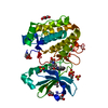


 PDBj
PDBj


 Assembly
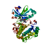

 SPODOPTERA FRUGIPERDA (fall armyworm) / References: UniProt: O15530, EC: 2.7.1.37
 Mass: 92.094 Da / Num. of mol.: 3 / Source method: obtained synthetically / Formula: C3H8O3
 Mass: 96.063 Da / Num. of mol.: 5 / Source method: obtained synthetically / Formula: SO4
 Mass: 466.531 Da / Num. of mol.: 1 / Source method: obtained synthetically / Formula: C28H26N4O3 / Comment: anticancer, antifungal, antibiotic, alkaloid*YM
 Mass: 18.015 Da / Num. of mol.: 97 / Source method: isolated from a natural source / Formula: H2O
 Sample preparation
 / Beamline: ID14-4 / Wavelength: 0.933
  Processing