 Movie
Movie Controller

[English] 日本語
 Yorodumi
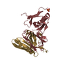
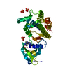
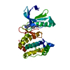
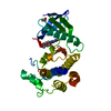
- PDB-1fpu: CRYSTAL STRUCTURE OF ABL KINASE DOMAIN IN COMPLEX WITH A SMALL MO... -
+ Open data

- Basic information
| Entry | Database: PDB / ID: 1fpu | ||||||
|---|---|---|---|---|---|---|---|
| Title | CRYSTAL STRUCTURE OF ABL KINASE DOMAIN IN COMPLEX WITH A SMALL MOLECULE INHIBITOR | ||||||
 Components Components | PROTO-ONCOGENE TYROSINE-PROTEIN KINASE ABL | ||||||
 Keywords Keywords | TRANSFERASE / kinase / kinase inhibitor / STI-571 / activation loop | ||||||
| Function / homology |  Function and homology information Function and homology informationRole of ABL in ROBO-SLIT signaling / HDR through Single Strand Annealing (SSA) / RHO GTPases Activate WASPs and WAVEs / MLL4 and MLL3 complexes regulate expression of PPARG target genes in adipogenesis and hepatic steatosis / Cyclin D associated events in G1 / Turbulent (oscillatory, disturbed) flow shear stress activates signaling by PIEZO1 and integrins in endothelial cells / Recruitment and ATM-mediated phosphorylation of repair and signaling proteins at DNA double strand breaks / protein localization to cytoplasmic microtubule plus-end / DNA conformation change / DN4 thymocyte differentiation ...Role of ABL in ROBO-SLIT signaling / HDR through Single Strand Annealing (SSA) / RHO GTPases Activate WASPs and WAVEs / MLL4 and MLL3 complexes regulate expression of PPARG target genes in adipogenesis and hepatic steatosis / Cyclin D associated events in G1 / Turbulent (oscillatory, disturbed) flow shear stress activates signaling by PIEZO1 and integrins in endothelial cells / Recruitment and ATM-mediated phosphorylation of repair and signaling proteins at DNA double strand breaks / protein localization to cytoplasmic microtubule plus-end / DNA conformation change / DN4 thymocyte differentiation / response to epinephrine / phospholipase C-inhibiting G protein-coupled receptor signaling pathway / podocyte apoptotic process / transitional one stage B cell differentiation / regulation of postsynaptic specialization assembly / positive regulation of phospholipase C/protein kinase C signal transduction / regulation of modification of synaptic structure / delta-catenin binding / regulation of cellular senescence / cerebellum morphogenesis / RUNX1 regulates transcription of genes involved in differentiation of HSCs / neuroepithelial cell differentiation / B cell proliferation involved in immune response / positive regulation of Wnt signaling pathway, planar cell polarity pathway / positive regulation of extracellular matrix organization / Regulation of actin dynamics for phagocytic cup formation / microspike assembly / B-1 B cell homeostasis / neuropilin signaling pathway / neuropilin binding / regulation of extracellular matrix organization / Myogenesis / bubble DNA binding / positive regulation of establishment of T cell polarity / activated T cell proliferation / positive regulation of blood vessel branching / proline-rich region binding / negative regulation of mitotic cell cycle / circulatory system development / mitogen-activated protein kinase binding / regulation of Cdc42 protein signal transduction / syntaxin binding / positive regulation of dendrite development / regulation of axon extension / regulation of T cell differentiation / alpha-beta T cell differentiation / positive regulation of cell migration involved in sprouting angiogenesis / negative regulation of cell-cell adhesion / neuromuscular process controlling balance / positive regulation of osteoblast proliferation / platelet-derived growth factor receptor-beta signaling pathway / platelet-derived growth factor receptor signaling pathway / Bergmann glial cell differentiation / cell leading edge / B cell proliferation / regulation of microtubule polymerization / negative regulation of long-term synaptic potentiation / negative regulation of cellular senescence / myoblast proliferation / associative learning / positive regulation of focal adhesion assembly / negative regulation of BMP signaling pathway / ephrin receptor signaling pathway / positive regulation of vasoconstriction / cardiac muscle cell proliferation / cellular response to transforming growth factor beta stimulus / negative regulation of endothelial cell apoptotic process / positive regulation of T cell migration / endothelial cell migration / negative regulation of double-strand break repair via homologous recombination / positive regulation of interleukin-2 production / phagocytosis / ephrin receptor binding / spleen development / four-way junction DNA binding / ruffle / canonical NF-kappaB signal transduction / positive regulation of stress fiber assembly / phosphotyrosine residue binding / signal transduction in response to DNA damage / peptidyl-tyrosine phosphorylation / actin filament polymerization / positive regulation of substrate adhesion-dependent cell spreading / substrate adhesion-dependent cell spreading / positive regulation of mitotic cell cycle / positive regulation of endothelial cell migration / SH2 domain binding / response to endoplasmic reticulum stress / thymus development / protein kinase C binding / positive regulation of release of sequestered calcium ion into cytosol / integrin-mediated signaling pathway / protein serine/threonine kinase activator activity / B cell receptor signaling pathway / post-embryonic development / regulation of actin cytoskeleton organization / non-specific protein-tyrosine kinase / neural tube closure / non-membrane spanning protein tyrosine kinase activity / cell-cell adhesion Similarity search - Function | ||||||
| Biological species |  | ||||||
| Method |  X-RAY DIFFRACTION / SYNCHROTRON / Resolution: 2.4 Å X-RAY DIFFRACTION / SYNCHROTRON / Resolution: 2.4 Å | ||||||
 Authors Authors | Schindler, T. / Bornmann, W. / Pellicena, P. / Miller, W.T. / Clarkson, B. / Kuriyan, J. | ||||||
 Citation Citation | Journal: Science / Year: 2000 Title: Structural mechanism for STI-571 inhibition of abelson tyrosine kinase. Authors: Schindler, T. / Bornmann, W. / Pellicena, P. / Miller, W.T. / Clarkson, B. / Kuriyan, J. | ||||||
| History |
|
- Structure visualization
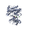
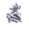
| Structure viewer | Molecule: MolmilJmol/JSmol |
|---|
- Downloads & links
-Download
| PDBx/mmCIF format | 1fpu.cif.gz | 122.8 KB | Display | PDBx/mmCIF format |
|---|---|---|---|---|
| PDB format | pdb1fpu.ent.gz | 95.2 KB | Display | PDB format |
| PDBx/mmJSON format | 1fpu.json.gz | Tree view | PDBx/mmJSON format | |
| Others |  Other downloads Other downloads |
-Validation report
| Arichive directory | https://data.pdbj.org/pub/pdb/validation_reports/fp/1fpuftp://data.pdbj.org/pub/pdb/validation_reports/fp/1fpu | HTTPS FTP |
|---|
-Related structure data
| Similar structure data |
|---|


-Links
 PDBj
PDBj


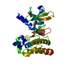
- Assembly
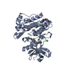
| Deposited unit | 
| ||||||||
|---|---|---|---|---|---|---|---|---|---|
| 1 | 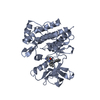
| ||||||||
| 2 | 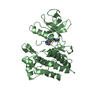
| ||||||||
| Unit cell |
| ||||||||
| Details | There are two kinase molecules in the asymmetric unit. The biological assembly is a monomer. |
-Components
| #1: Protein | Mass: 33743.523 Da / Num. of mol.: 2 / Fragment: KINASE DOMAIN Source method: isolated from a genetically manipulated source Source: (gene. exp.)   Spodoptera frugiperda (fall armyworm) / References: UniProt: P00520, EC: 2.7.1.112 Spodoptera frugiperda (fall armyworm) / References: UniProt: P00520, EC: 2.7.1.112#2: Chemical |   Mass: 382.418 Da / Num. of mol.: 2 / Fragment: 2-(PHENYLAMINO)-PYRIMIDINE COMPOUND / Source method: obtained synthetically / Formula: C22H18N6O Mass: 382.418 Da / Num. of mol.: 2 / Fragment: 2-(PHENYLAMINO)-PYRIMIDINE COMPOUND / Source method: obtained synthetically / Formula: C22H18N6ODetails: chemically synthesized as described in Zimmermann J., Buchdunger E., Mett H., Meyer T., Lydon N.B. (1997) Bioorg. Med. Chem. Lett., Vol. 7, pp. 187 #3: Water | ChemComp-HOH / |  Mass: 18.015 Da / Num. of mol.: 99 / Source method: isolated from a natural source / Formula: H2O Mass: 18.015 Da / Num. of mol.: 99 / Source method: isolated from a natural source / Formula: H2O |
|---|
-Experimental details
-Experiment
| Experiment | Method: X-RAY DIFFRACTION / Number of used crystals: 1 |
|---|
- Sample preparation
| Crystal | Density Matthews: 2.29 Å3/Da / Density % sol: 46.24 % | ||||||||||||||||||||||||||||||||||||||||||||||||
|---|---|---|---|---|---|---|---|---|---|---|---|---|---|---|---|---|---|---|---|---|---|---|---|---|---|---|---|---|---|---|---|---|---|---|---|---|---|---|---|---|---|---|---|---|---|---|---|---|---|
| Crystal grow | Temperature: 277 K / Method: vapor diffusion, hanging drop / pH: 6.2 Details: PEG 4000, magnesium chloride, mes, pH 6.2, VAPOR DIFFUSION, HANGING DROP, temperature 277K | ||||||||||||||||||||||||||||||||||||||||||||||||
| Crystal grow | *PLUS Temperature: 4 ℃ / pH: 8 | ||||||||||||||||||||||||||||||||||||||||||||||||
| Components of the solutions | *PLUS
|
-Data collection
| Diffraction | Mean temperature: 103 K |
|---|---|
| Diffraction source | Source: SYNCHROTRON / Site: NSLS  / Beamline: X25 / Wavelength: 0.9393 / Beamline: X25 / Wavelength: 0.9393 |
| Detector | Type: BRANDEIS - B4 / Detector: CCD / Date: Sep 5, 1999 |
| Radiation | Protocol: SINGLE WAVELENGTH / Monochromatic (M) / Laue (L): M / Scattering type: x-ray |
| Radiation wavelength | Wavelength: 0.9393 Å / Relative weight: 1 |
| Reflection | Resolution: 2.4→99 Å / Num. obs: 221636 / % possible obs: 98.7 % / Redundancy: 9.3 % / Biso Wilson estimate: 61 Å2 / Rmerge(I) obs: 0.068 / Net I/σ(I): 30.9 |
| Reflection shell | Resolution: 2.4→2.49 Å / Redundancy: 5.7 % / Rmerge(I) obs: 0.216 / Num. unique all: 2190 / % possible all: 90.6 |
| Reflection | *PLUS Num. obs: 24122 / Num. measured all: 221636 |
| Reflection shell | *PLUS % possible obs: 90.6 % |
- Processing
| Software |
| |||||||||||||||||||||||||
|---|---|---|---|---|---|---|---|---|---|---|---|---|---|---|---|---|---|---|---|---|---|---|---|---|---|---|
| Refinement | Resolution: 2.4→99 Å / Stereochemistry target values: Engh & Huber Details: Tight, noncrystallographic restraints were utilized throughout the refinement so that the final r.m.s. deviation between the two molecules is 0.05 and 0.04 Angstrom for the N-terminal and ...Details: Tight, noncrystallographic restraints were utilized throughout the refinement so that the final r.m.s. deviation between the two molecules is 0.05 and 0.04 Angstrom for the N-terminal and the C-terminal lobes, respectively (excluding residues 229 to 337, 252, 262, 271, 294, 306, 404, 447, 450, 466, and 491, which were built in different conformations in the two molecules). The electron density map was significantly weaker for one of the two molecules in the asymmetric unit (chain ID B), and all the analysis (cf. primary citation) relied on the better-ordered one (chain ID A).
| |||||||||||||||||||||||||
| Refinement step | Cycle: LAST / Resolution: 2.4→99 Å
| |||||||||||||||||||||||||
| Refine LS restraints |
| |||||||||||||||||||||||||
| Software | *PLUS Name: CNS / Classification: refinement | |||||||||||||||||||||||||
| Refinement | *PLUS Highest resolution: 2.4 Å / Lowest resolution: 99 Å / Rfactor obs: 0.239 | |||||||||||||||||||||||||
| Solvent computation | *PLUS | |||||||||||||||||||||||||
| Displacement parameters | *PLUS | |||||||||||||||||||||||||
| Refine LS restraints | *PLUS Type: c_angle_deg / Dev ideal: 1.4 |
