 Movie
Movie Controller


+ Open data

- Basic information
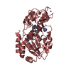
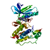
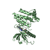
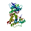
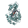
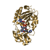
| Entry | Database: PDB / ID: 4c78 | ||||||
|---|---|---|---|---|---|---|---|
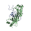
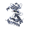
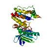
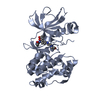
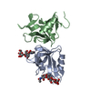
| Title | Complex of human Sirt3 with Bromo-Resveratrol and ACS2 peptide | ||||||
 Components Components |
| ||||||
 Keywords Keywords | HYDROLASE / SIRTUIN / INHIBITOR / ACTIVATION / RESVERATROL / SIRT1 / METABOLIC SENSOR / METABOLISM / AGING | ||||||
| Function / homology |  Function and homology information Function and homology informationpropionate biosynthetic process / propionate-CoA ligase / acetate biosynthetic process / propionate-CoA ligase activity / positive regulation of catalase activity / positive regulation of superoxide dismutase activity / NAD-dependent protein lysine delactylase activity / positive regulation of ceramide biosynthetic process / acetate-CoA ligase / acetyl-CoA synthetase activity ...propionate biosynthetic process / propionate-CoA ligase / acetate biosynthetic process / propionate-CoA ligase activity / positive regulation of catalase activity / positive regulation of superoxide dismutase activity / NAD-dependent protein lysine delactylase activity / positive regulation of ceramide biosynthetic process / acetate-CoA ligase / acetyl-CoA synthetase activity / : / ethanol catabolic process / Ethanol oxidation / peptidyl-lysine deacetylation / Maturation of TCA enzymes and regulation of TCA cycle / acetyl-CoA biosynthetic process / protein acetyllysine N-acetyltransferase / protein deacetylation / NAD-dependent protein lysine deacetylase activity / histone deacetylase activity, NAD-dependent / positive regulation of oxidative phosphorylation / Regulation of FOXO transcriptional activity by acetylation / protein lysine deacetylase activity / AMP binding / cellular response to stress / negative regulation of reactive oxygen species metabolic process / NAD+ binding / Mitochondrial unfolded protein response (UPRmt) / FOXO-mediated transcription of oxidative stress, metabolic and neuronal genes / Transferases; Acyltransferases; Transferring groups other than aminoacyl groups / aerobic respiration / Transcriptional activation of mitochondrial biogenesis / negative regulation of ERK1 and ERK2 cascade / positive regulation of insulin secretion / sequence-specific DNA binding / mitochondrial matrix / enzyme binding / protein-containing complex / mitochondrion / zinc ion binding / nucleoplasm / ATP binding / nucleus Similarity search - Function | ||||||
| Biological species |  HOMO SAPIENS (human) HOMO SAPIENS (human) | ||||||
| Method |  X-RAY DIFFRACTION / SYNCHROTRON / MOLECULAR REPLACEMENT / Resolution: 2 Å X-RAY DIFFRACTION / SYNCHROTRON / MOLECULAR REPLACEMENT / Resolution: 2 Å | ||||||
 Authors Authors | Nguyen, G.T.T. / Gertz, M. / Weyand, M. / Steegborn, C. | ||||||
 Citation Citation | Journal: Chem.Biol. / Year: 2013 Title: Crystal Structures of Sirt3 Complexes with 4'-Bromo-Resveratrol Reveal Binding Sites and Inhibition Mechanism. Authors: Nguyen, G.T.T. / Gertz, M. / Steegborn, C. | ||||||
| History |
|
- Structure visualization
| Structure viewer | Molecule: MolmilJmol/JSmol |
|---|
- Downloads & links
-Download
| PDBx/mmCIF format | 4c78.cif.gz | 125.5 KB | Display | PDBx/mmCIF format |
|---|---|---|---|---|
| PDB format | pdb4c78.ent.gz | 97.5 KB | Display | PDB format |
| PDBx/mmJSON format | 4c78.json.gz | Tree view | PDBx/mmJSON format | |
| Others |  Other downloads Other downloads |
-Validation report
| Arichive directory | https://data.pdbj.org/pub/pdb/validation_reports/c7/4c78ftp://data.pdbj.org/pub/pdb/validation_reports/c7/4c78 | HTTPS FTP |
|---|
-Related structure data
| Related structure data | 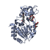 4c7bC  4h8d C: citing same article ( S: Starting model for refinement |
|---|---|
| Similar structure data |

-Links
 PDBj
PDBj


- Assembly
| Deposited unit | 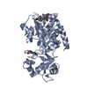
| ||||||||
|---|---|---|---|---|---|---|---|---|---|
| 1 |
| ||||||||
| Unit cell |
| ||||||||
| Components on special symmetry positions |
|
-Components
| #1: Protein | Mass: 31484.219 Da / Num. of mol.: 1 / Fragment: RESIDUES 116-399 Source method: isolated from a genetically manipulated source Source: (gene. exp.) HOMO SAPIENS (human) / Plasmid: PVFT3S / Production host:  References: UniProt: Q9NTG7, Hydrolases; Acting on carbon-nitrogen bonds, other than peptide bonds; In linear amides |
|---|---|
| #2: Protein/peptide | Mass: 1248.523 Da / Num. of mol.: 1 / Fragment: RESIDUES 638-647 / Source method: obtained synthetically / Source: (synth.) HOMO SAPIENS (human) / References: UniProt: Q9NUB1, acetate-CoA ligase |
| #3: Chemical | ChemComp-BVB /   Mass: 291.140 Da / Num. of mol.: 1 / Source method: obtained synthetically / Formula: C14H11BrO2 Mass: 291.140 Da / Num. of mol.: 1 / Source method: obtained synthetically / Formula: C14H11BrO2 |
| #4: Chemical | ChemComp-ZN /   Mass: 65.409 Da / Num. of mol.: 1 / Source method: obtained synthetically / Formula: Zn Mass: 65.409 Da / Num. of mol.: 1 / Source method: obtained synthetically / Formula: Zn |
| #5: Water | ChemComp-HOH /  Mass: 18.015 Da / Num. of mol.: 125 / Source method: isolated from a natural source / Formula: H2O Mass: 18.015 Da / Num. of mol.: 125 / Source method: isolated from a natural source / Formula: H2O |
| Has protein modification | Y |
-Experimental details
-Experiment
| Experiment | Method: X-RAY DIFFRACTION / Number of used crystals: 1 |
|---|
- Sample preparation
| Crystal | Density Matthews: 2.33 Å3/Da / Density % sol: 47.2 % / Description: NONE |
|---|---|
| Crystal grow | pH: 6 Details: 250 MM (NH4)2SO4, 100 MM BISTRIS PH 6, 21% (W/V) PEG 3350 |
-Data collection
| Diffraction | Mean temperature: 100 K |
|---|---|
| Diffraction source | Source: SYNCHROTRON / Site: BESSY  / Beamline: 14.1 / Wavelength: 0.918 / Beamline: 14.1 / Wavelength: 0.918 |
| Detector | Type: MARMOSAIC 225 mm CCD / Detector: CCD / Date: Nov 29, 2011 / Details: COLLIMATOR |
| Radiation | Monochromator: SI(111) MONOCHROMATOR / Protocol: SINGLE WAVELENGTH / Monochromatic (M) / Laue (L): M / Scattering type: x-ray |
| Radiation wavelength | Wavelength: 0.918 Å / Relative weight: 1 |
| Reflection | Resolution: 2→37.4 Å / Num. obs: 37699 / % possible obs: 98.6 % / Observed criterion σ(I): -3 / Redundancy: 2.1 % / Rmerge(I) obs: 0.05 / Net I/σ(I): 11.5 |
| Reflection shell | Resolution: 2→2.1 Å / Redundancy: 2.1 % / Rmerge(I) obs: 0.43 / Mean I/σ(I) obs: 1.99 / % possible all: 99 |
- Processing
| Software |
| ||||||||||||||||||||||||||||||||||||||||||||||||||||||||||||||||||||||||||||||||||||||||||||||||||||||||||||||||||||||||||||||||||||||||||||||||||||||||||||||||||||||||||||||||||||||
|---|---|---|---|---|---|---|---|---|---|---|---|---|---|---|---|---|---|---|---|---|---|---|---|---|---|---|---|---|---|---|---|---|---|---|---|---|---|---|---|---|---|---|---|---|---|---|---|---|---|---|---|---|---|---|---|---|---|---|---|---|---|---|---|---|---|---|---|---|---|---|---|---|---|---|---|---|---|---|---|---|---|---|---|---|---|---|---|---|---|---|---|---|---|---|---|---|---|---|---|---|---|---|---|---|---|---|---|---|---|---|---|---|---|---|---|---|---|---|---|---|---|---|---|---|---|---|---|---|---|---|---|---|---|---|---|---|---|---|---|---|---|---|---|---|---|---|---|---|---|---|---|---|---|---|---|---|---|---|---|---|---|---|---|---|---|---|---|---|---|---|---|---|---|---|---|---|---|---|---|---|---|---|---|
| Refinement | Method to determine structure: MOLECULAR REPLACEMENT Starting model: PDB ENTRY 4H8D 4h8d Resolution: 2→37.42 Å / Cor.coef. Fo:Fc: 0.957 / Cor.coef. Fo:Fc free: 0.939 / SU B: 9.553 / SU ML: 0.137 / Cross valid method: THROUGHOUT / ESU R: 0.184 / ESU R Free: 0.167 / Stereochemistry target values: MAXIMUM LIKELIHOOD Details: HYDROGENS HAVE BEEN ADDED IN THE RIDING POSITIONS. U VALUES WITH TLS ADDED.
| ||||||||||||||||||||||||||||||||||||||||||||||||||||||||||||||||||||||||||||||||||||||||||||||||||||||||||||||||||||||||||||||||||||||||||||||||||||||||||||||||||||||||||||||||||||||
| Solvent computation | Ion probe radii: 0.8 Å / Shrinkage radii: 0.8 Å / VDW probe radii: 1.2 Å / Solvent model: MASK | ||||||||||||||||||||||||||||||||||||||||||||||||||||||||||||||||||||||||||||||||||||||||||||||||||||||||||||||||||||||||||||||||||||||||||||||||||||||||||||||||||||||||||||||||||||||
| Displacement parameters | Biso mean: 40.552 Å2
| ||||||||||||||||||||||||||||||||||||||||||||||||||||||||||||||||||||||||||||||||||||||||||||||||||||||||||||||||||||||||||||||||||||||||||||||||||||||||||||||||||||||||||||||||||||||
| Refinement step | Cycle: LAST / Resolution: 2→37.42 Å
| ||||||||||||||||||||||||||||||||||||||||||||||||||||||||||||||||||||||||||||||||||||||||||||||||||||||||||||||||||||||||||||||||||||||||||||||||||||||||||||||||||||||||||||||||||||||
| Refine LS restraints |
|